Article

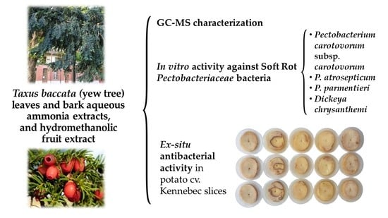
Phytochemical Screening and Antibacterial Activity of Taxus baccata L. against Pectobacterium spp. and Dickeya chrysanthemi

by
Eva Sánchez-Hernández
1,
Vicente González-García
2,
Jesús Martín-Gil
1,
Belén Lorenzo-Vidal
3,
Ana Palacio-Bielsa
2 and
Pablo Martín-Ramos
1,*
1
Department of Agricultural and Forestry Engineering, ETSIIAA, Universidad de Valladolid, Avenida de Madrid 44, 34004 Palencia, Spain
2
Department of Agricultural, Forest and Environmental Systems, Agrifood Research and Technology Centre of Aragón, Instituto Agroalimentario de Aragón—IA2 (Universidad de Zaragoza-CITA), Avda. Montañana 930, 50059 Zaragoza, Spain
3
Servicio de Microbiología, Hospital Universitario Rio Hortega, Calle Dulzaina 2, 47012 Valladolid, Spain
*
Author to whom correspondence should be addressed.
Horticulturae 2023, 9(2), 201; https://doi.org/10.3390/horticulturae9020201
Submission received: 29 December 2022 / Revised: 27 January 2023 / Accepted: 31 January 2023 / Published: 3 February 2023

Abstract

:
The yew tree (Taxus baccata L.) is considered in folklore a symbol of immortality due to its qualities of longevity and regeneration. Despite its poisonous reputation, the yew tree has a long history of medicinal use, particularly in the form of extracts from its leaves and bark. In the work presented herein, gas chromatography–mass spectrometry (GC–MS) chemical profiling was applied to the aqueous ammonia/hydromethanolic extracts of several plant organs of T. baccata, leading to the identification of different bioactive compounds than those previously characterized by high-performance liquid chromatography with tandem mass spectrometry (HPLC–MS/MS) in other extraction media. The leaf aqueous ammonia extract was rich in 2-hexylthiophene and 3-O-methyl-d-fructose; 9-octadecenoic and hexadecanoic acid were the main constituents of the bark aqueous ammonia extract; and the fruit hydromethanolic extract contained methyl 2-O-methyl-α-d-xylofuranoside, 1,3-dioxolane derivatives, and erysimoside. The antimicrobial activity of the extracts was assayed against four bacterial pathogens responsible for the soft rot and blackleg diseases of potatoes, viz. Pectobacterium carotovorum subsp. carotovorum, Pectobacterium atrosepticum, Pectobacterium parmentieri, and Dickeya chrysanthemi, resulting in minimum inhibitory concentration (MIC) values as low as 187 μg·mL−1. Bioassays on potato slices confirmed the efficacy of the leaf extract at this dose when applied as a preventive treatment before artificial inoculation with P. carotovorum subsp. carotovorum. In view of this high activity, these extracts may find application in the integrated pest management of soft rot Pectobacteriaceae (SRP) diseases.

Graphical Abstract

1. Introduction

The yew (Taxus baccata L.) is an evergreen coniferous tree in the Taxaceae family that is native to Western, Central, and Southern Europe, Northwestern Africa, and Southwest Asia. It grows in mixed forests, preferentially in shady areas, and is indifferent about its substrate. It is mainly grown for ornamental purposes. Yews are small to medium-sized trees that grow 10–20 m tall and have trunks up to 2 m (rarely 4 m) in diameter. The bark is thin and scaly brown, and it peels off in small flakes parallel to the stem. The leaves are flat, dark green, 1–4 cm long, and 2–3 mm wide. They are arranged in a spiral on the stem, although the leaf bases are twisted to line the leaves in two flat rows on one and the other side of the stem. The seeds are 6–7 mm long and have a woody episperm that is mostly covered by a fleshy red aril. The yew is dioecious, so the male and female flowers grow on different trees.
The yew tree has a long history of medicinal use, particularly in the form of extracts from its leaves and bark. Concerning its medicinal uses in folklore, the leaves and stem are anti-inflammatory, antirheumatic, abortifacient, purgative, and diaphoretic, and were traditionally used for epilepsy, malaria, rheumatism, tuberculosis, cold, and coughs; a tincture from young shoots was used to treat headaches, diarrhea, biliousness, and giddiness; the bark was used to treat coughs and colds; and pounded leaves were administered orally for indigestion, asthma, and bronchial problems [1]. Nonetheless, except for the red arils, scientific data indicate that the plant’s remaining parts are poisonous, including toxins that can be absorbed through skin contact and inhalation. The consumption of even a small amount of leaves, seeds, or bark can result in death. Selected compounds, such as baccatin III; 10-deacetylbaccatin III (10-DAB III); 3,5-dimethoxyphenol; paclitaxel (taxol A); cephalomannine (taxol B); 10-deacetylpaclitaxel; 10-deacetylcephalomannine; taxcultine; epicanadensene; and taxinine M were identified by high-performance liquid chromatography with tandem mass spectrometry (HPLC–MS/MS) method [2,3,4]. All are very toxic [2]. However, mature ripe fruits are free of toxic compounds [5,6,7,8].
Yew tree extracts have garnered significant attention as a potential treatment for cancer, given that taxol and related taxanes (e.g., docetaxel or Taxotere®) have been shown to inhibit cancer cell growth and trigger apoptosis in a variety of cancer types [9]. However, in spite of the fact that T. baccata is quite resistant to decay as well as to attacks of parasites and pests, pointing to the existence of bioactive compounds with potential applications in crop protection, its extracts have only been assayed against the red spider mite (Tetranychus urticae Koch) [10].
Concerning crop protection, a particularly interesting application would be the control of tuber soft rot caused by Pectobacteriaceae (SRP), which is among the most important potato (Solanum tuberosum L.) diseases, both for seed and ware potato production, resulting in overall losses estimated at 46 M euro/year for the European sector (with high variability among years) [11], because of the absence of authorized and effective crop protection chemicals against SRP [12], and, at least for now, lack of disease resistance. SRP can result in severe losses in important economical crops in many nations. It affects a wide host range, including three of the four most important crops in the world (viz. rice, maize, and potato).
The most common sign of potato blackleg is a slimy, moist, black rot lesion that spreads up the stems from the rotting mother tuber, particularly under wet conditions. Under dry conditions, symptoms include the stunting, wilting, yellowing, and desiccation of the leaves and stems [13]. Tuber soft rot begins at the stolon end, lenticels, and/or in wounds. Blanking happens when seed tubers start rotting in the field prior to emergence. The tuber tissue is macerated to a creamy consistency; in the presence of air, it turns black and has an unpleasant odor when secondary organisms invade it. Rotting can spread to neighboring tubers in poorly ventilated cool storage [14].
The aim of the study presented herein was twofold: (i) to investigate the constituents of needle and bark aqueous ammonia extracts, as well as of a red aril hydromethanolic extract, using gas chromatography–mass spectrometry (GC−MS) in order to analyze the influence of the extraction media and quantification methods on the presence/absence of the aforementioned phytochemicals; and (ii) to assay the antimicrobial activity of the extracts against four potato SRP species ranked in the top ten plant pathogens in 2010 [15], viz. Pectobacterium carotovorum subsp. carotovorum (Jones 1901) Hauben et al. 1999; Pectobacterium atrosepticum (van Hall 1902) Gardan et al. 2003; Pectobacterium parmentieri Khayi et al. 2016; and Dickeya chrysanthemi (Burkholder et al. 1953) Samson et al. 2005. The reported findings may also be applied to other vegetable crops, such as chicory and radish, which have also suffered severe soft rot epidemics in the past decades.

2. Material and Methods

2.1. Plant Material and Reagents

Samples of needle leaves, bark, and mature fruits were collected from a centenarian yew (female tree) growing in an ancient square (the Viejo Coso, i.e., Old Bullring [tr.]; 41°39′23.7″ N 4°43′42.0″ W) in Valladolid, Spain. Sampling took place on 20 September 2022.
Ammonium hydroxide, 50% v/v aq. soln. (CAS 1336-21-6), acetic acid (CAS 64-19-7; glacial, ≥99%), tryptic soy agar (TSA, CAS 91079-40-2), and tryptic soy broth (TSB, CAS 8013-01-2) were purchased from Sigma-Aldrich Química (Madrid, Spain).

2.2. Bacterial Strains

Pectobacterium carotovorum subsp. carotovorum (strain CITA Ecc-8), Pectobacterium atrosepticum (strain CITA Eca-2), and Dickeya chrysanthemi (strain CIRM-CFBP 7086) were supplied by the Bacteriology Laboratory at the Center for Research and Agrifood Technology of Aragón (CITA, Zaragoza, Spain). Pectobacterium parmentieri (strain CRD 16/111) was obtained from the Regional Diagnostic Center of the Junta de Castilla y León (Salamanca, Spain). All of these bacterial strains were isolated from diseased potatoes in Spain. The bacterial strains were cultured on TSA medium.

2.3. Extracts Preparation

Concerning the choice of the extraction media, the hydroalcoholic medium—a very popular option when characterization is to be conducted by GC−MS [16]—was chosen for the arils. However, it does not allow for the dissolution of polyphenols and other bioactive compounds of interest contained in the bark samples, which can be successfully attained by digestion in an aqueous ammonia solution (widely used in lignocellulosic residue pretreatment [17]), as previously shown in other works involving bark extracts [18,19,20]. Concerning the leaf extraction medium, aqueous ammonia was chosen due to the substantially higher extraction yield in comparison with the hydromethanolic medium (an order of magnitude higher), probably due to the high lignin content (avg. value of 18.28 ± 5.55%, according to [21]) of yew needles.
The preparation of the bark and leaf extracts was performed according to the procedure described in [22] with the modifications indicated in [20]. In brief, the powdered samples (31 g) were digested in an aqueous ammonia solution (120 mL H2O + 30 mL NH3(aq.)) for 2 h before being sonicated for 10 min in pulsed mode (with a 2 min stop every 2.5 min) with a model UIP1000hdT probe-type ultrasonicator (Hielscher Ultrasonics, Teltow, Germany), neutralized with acetic acid, and allowed to stand for 24 h. After centrifuging the solution at 9000 rpm for 15 min, the supernatants were filtered using Whatman No. 1 paper. The preparation of the fruit (red arils) hydroalcoholic extract followed the same procedure, albeit in a hydromethanolic medium (100 g in 250 mL of MeOH:H2O, 1:1 v/v) and without the neutralization step.
The samples of the extracts were freeze-dried to obtain the solid residue. The extraction yields were 1.92%, 11.15%, and 11.3% for the bark, leaf, and fruit extracts, respectively. For subsequent GC−MS analysis, 25 mg of the obtained freeze-dried extracts were dissolved in 5 mL of HPLC-grade methanol to acquire a 5 mg·mL−1 solution, which was then filtered.

2.4. Extracts Characterization

The infrared spectra of the dried plant organs and their freeze-dried extracts were recorded using a Nicolet iS50 Fourier-transform infrared spectrometer (Thermo Scientific; Waltham, MA, USA) with an in-built diamond attenuated total reflectance system. The spectra were registered spanning the 400–4000 cm−1 range, with a spectral resolution of 1 cm−1, using the interferograms produced by co-adding 64 scans.
The extracts were further studied by GC−MS at Universidad de Alicante’s Research Support Services (STI) using a model 7890A gas chromatograph coupled to a model 5975C quadrupole mass spectrometer (Agilent Technologies; Santa Clara, CA, USA). The following chromatographic conditions were used: 3 injections/vial, injection volume = 1 µL; injector temperature = 280 °C, in splitless mode; initial oven temperature = 60 °C, after 2 min, followed by a 10 °C·min−1 increase up to a final temperature of 300 °C after 15 min. The chromatographic column utilized for compound separation was an HP-5MS UI (Agilent Technologies) of 30 m in length, 0.250 mm in diameter, and with 0.25 µm film. The mass spectrometer was configured as follows: temperature of the mass spectrometer’s electron impact source = 230 °C; temperature of the quadrupole = 150 °C; ionization energy = 70 eV. For equipment calibration, test mixture 2 for apolar capillary columns according to Grob (https://www.sigmaaldrich.com/ES/es/product/sial/86501, accessed on 29 January 2023) and PFTBA were utilized as tuning standards. The components were identified by comparing their mass spectra and retention times with those of authentic compounds, as well as by computer matching with the National Institute of Standards and Technology (NIST, Gaithersburg, MD, USA) database.

2.5. In Vitro Antimicrobial Activity Assessment

The antibacterial activity of the extracts was assessed using the agar dilution technique in accordance with the Clinical and Laboratory Standards Institute (CLSI, Wayne, PA, USA) standard M07–11 [23], determining the minimum inhibitory concentrations (MIC). Pure cultures of each Pectobacterium and Dickeya species were incubated in a TSB liquid medium for 24 h at 28 °C. Following that, serial dilutions were performed, beginning with a concentration of 108 CFU·mL−1 and ending with a final inoculum of ~104 CFU·mL−1. The bacterial suspensions were then spread on the surface of the TSA plates, to which the bioactive products had previously been added at concentrations in the 62.5 to 1500 μg·mL−1 range. After 24 h of incubation at 28 °C, bacterial growth was recorded. The MICs were determined as the lowest bioactive product concentrations at which no bacterial growth was observed. All experiments were run in triplicate, with three plates for each treatment and concentration combination.
For comparison purposes, MIC values for five conventional antibiotics of clinical use (viz. benzylpenicillin or penicillin G (PG); ampicillin (AM); gentamicin (GM); ciprofloxacin (CI); and tetracycline (TC)) were determined using ETEST® strips.

2.6. Protection Tests on Potato Slices

The potato tubers (Solanum tuberosum cv. Kennebec) used to determine the protective and curative effect of T. baccata leaf extract were supplied by M. Prado Mazaira S.L. (Coristanco, A Coruña, Spain) and were previously cultivated according to organic farming regulations, with no use of synthetic pesticides, under the ‘Pataca de Galicia’ protected geographical indication. The tubers were harvested and immediately cold shipped by an express courier service so that experiments could be started within 24 h after harvesting. Tubers were selected based on uniformity of size, with a caliber ≥45 mm and an absence of physical damage and fungal or bacterial infection symptoms.
The efficacy of the treatment was determined by artificially inoculating potato slices in controlled laboratory conditions. Inoculation was carried out in accordance with the method proposed by Abd-El-Khair et al. [24]. Briefly, the potato tubers were washed under running water to remove soil and other organic particles before being superficially disinfected for 2 min with a NaOCl 3% solution, washed three times with sterile distilled water, and dried in a laminar flow hood on sterile absorbent paper. Then, under sterile conditions, potato tubers were cut into 8 mm-thick slices with a sterile knife. In each Petri plate containing sterile filter paper soaked with 3 mL of sterile distilled water, one potato slice was placed. Then, a superficial wound (ø = 3 mm) was created in the equatorial zone of each slice, where 50 µL of the bacterial suspension (104 CFU·mL−1) of P. carotovorum subsp. carotovorum strain CITA Ecc-8 was inoculated simultaneously with the addition of 50 µL of T. baccata leaf extract at a concentration of 187.5 µg·mL−1 (i.e., the MIC obtained in previous in vitro assays). The same procedure was repeated, but successively, applying the extract either 2 h before or after pathogen inoculation. The inoculated potato slices were incubated at 30 °C ± 2 for 48 h. For negative controls, potato slices were treated with distilled water. Three independent trials were carried out, and each trial consisted of three replicate potato slices per treatment, in agreement with Garge and Nerurkar [25]. The macerated tissue was removed and weighed in order to calculate the relative percentage maceration of the potato slices, using the positive control as 100%, according to the procedure described in [26]. The results of the relative percentage maceration were statistically analyzed in IBM SPSS Statistics v.25 software (IBM, Armonk, NY, USA) by one-way analysis of variance, followed by a post hoc comparison of means by Tukey’s test (because the requirements of homogeneity and homoscedasticity were met, according to the Shapiro–Wilk and Levene tests).

3. Results

3.1. Vibrational Characterization

The main bands in the infrared spectra of the different plant organs and their extracts are summarized in Table 1. The presence of thiophene rings would be supported by the aromatic C−C ring stretching at 1652 and 1374 cm−1 and the peak observed at 720 cm−1 for C−S bending. The bands at ca. 1460, 1245, 790, and 779 cm−1 show the connectivity of thiophene rings through 2,5 coupling [27]. The bands at 2849, 1409, 1255, and 1193 cm−1 have been previously reported for methyl d-lyxopyranosides [28]; those at 1143, 1023, 918, and 865 cm−1 for dioxolanes [29]; and those at 2915, 1698, and 1102 cm−1 for hydroxy-cyclopentenones [30].

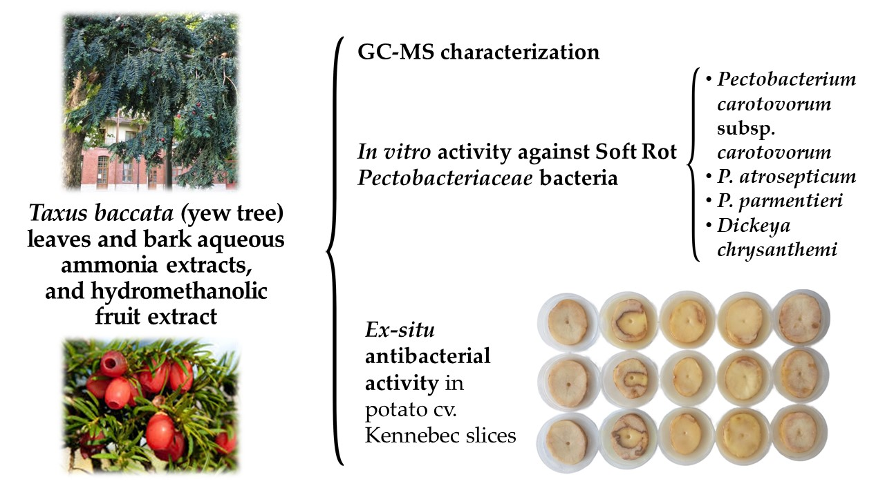
3.2. GC–MS Chromatograms

The main constituents identified in the leaf aqueous ammonia extract (Figure 1 and Figure S1, Table S1) were: 2-hexylthiophene (22.4%); 3-O-methyl-d-fructose (16.1%); catechol (6.2%); 2,3-dimethyl-cyclohexa−1,3-diene (7.7%); phenol (6.9%) and phenol derivatives (5.8%); 2-methyl−1,3-cyclohexanedione (4.1%); 2-cyclopenten-1-one derivatives (4.1%); and 2,3-dihydro-benzofurane (3.4%).
In the bark aqueous ammonia extract (Figure 1 and Figure S2, Table S2), the following phytochemicals were detected: 9-octadecenoic acid, methyl ester (15.7%); hexadecanoic acid, methyl ester (13.5%); undecane (12.2%); 4-acetamidobenzofuroxane (11.7%); methoxy-phenyl-oxime (11.3%); 3,4-methylenedioxyphenyl acetone (8.1%); catechol (6.4%); methyl stearate (4.8%); 1-methyl-4-(1-methylpropyl)-benzene (4.2%); dodecanoic acid methyl ester (3.5%); and 1-cyclohexene-1-carboxylic acid (3.2%).
In turn, in the fruit hydromethanolic extract (Figure 1 and Figure S3, Table S3), GC–MS pointed to the presence of 5-hydroxymethylfurfural (5-HMF, 13.3%) and furfural (1.1%); methyl 2-O-methyl-α-d-xylofuranoside (8.3%); 1,3-dioxolane derivatives (8.2%); 3-deoxy-d-mannoic lactone (5.9%); 2,3-dihydro-3,5-dihydroxy-6-methyl-4H-pyran-4-one (5.5%); 3-[(4-nitrobutyl)thio]−1-propene (5.3%); dihydroxyacetone (3.3%); 2,2-dimethyl-3-oxo-butanoic acid methyl ester (2.4%); 1,4-dinitroso piperazine (1.6%); 2,3-dihydroxy propanal; 2,4-dihydroxy-2,5-dimethyl-3(2H)-furan-3-one (1.5%); 2-ethyltetrahydro-thiophene (1.3%); erysimoside or (3β,5β)-3-{[2,6-dideoxy-4-O-(β-d-glucopyranosyl)-β-d-ribo-hexopyranosyl]oxy}-5,14-dihydroxy−19-oxocard-20(22)-enolide (1.0%); 2-hydroxy-2-cyclopenten-1-one (0.7%); tetrahydro-2-methylthiophene (0.9%); 2-hexyl thiophene (0.8%); and catechol (0.3%).

3.3. Antibacterial Activity of the Extracts

3.3.1. In vitro Assays

Table 2 summarizes the antibacterial activity results against the four SRP taxa. The hydromethanolic fruit extract was the least effective in all cases, with MIC values of 1000–1500 μg·mL−1; the bark aqueous ammonia extract showed an intermediate activity, with MIC values ranging from 500 to 750 μg·mL−1; whereas the leaf aqueous ammonia extract resulted in the highest inhibition, with MIC values of 187.5 μg·mL−1 against all bacterial strains.
MIC values for five conventional antibiotics (viz. benzylpenicillin, ampicillin, gentamicin, ciprofloxacin, and tetracycline), determined using ETEST® strips (Figure S4), are provided in Table 3 for comparison purposes.

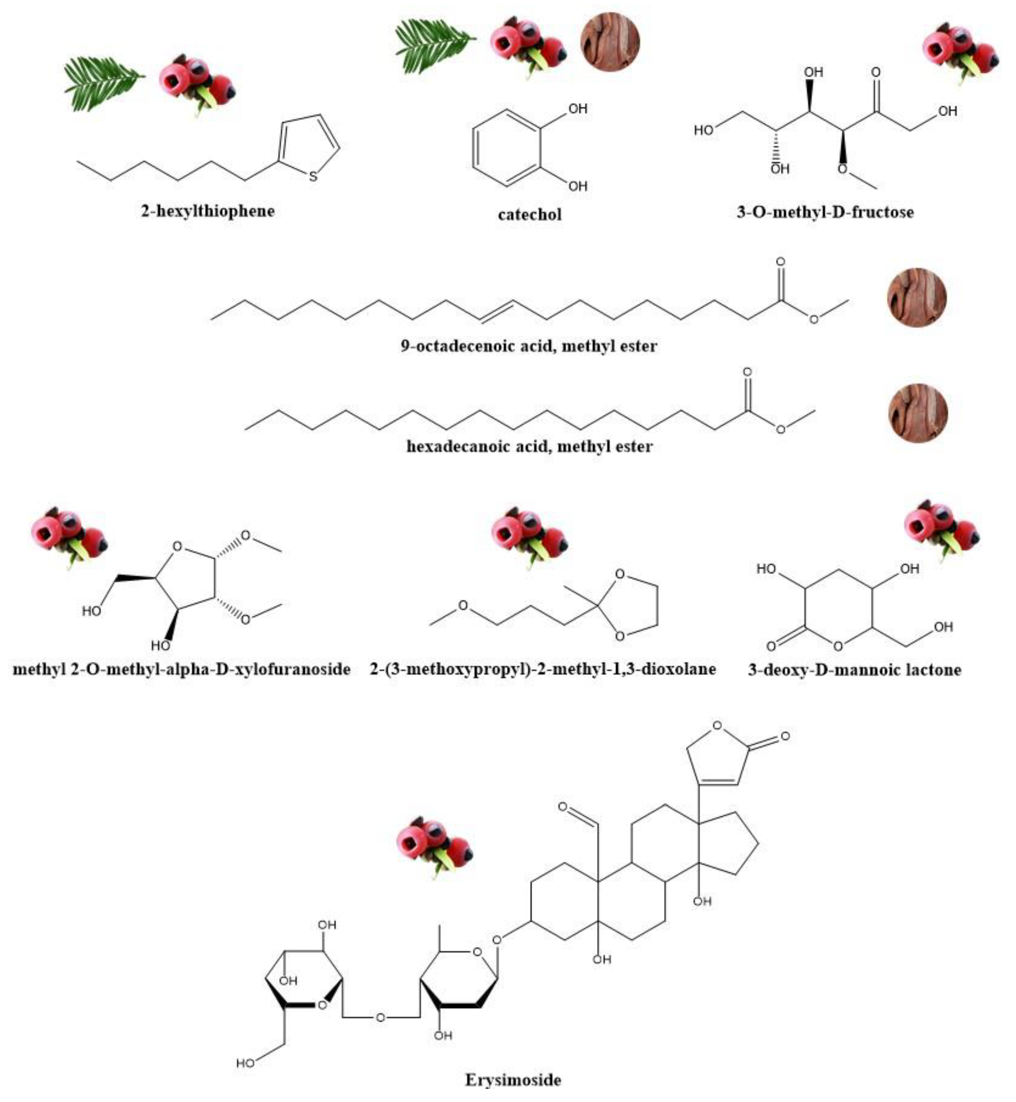
3.3.2. Bioassays on Potato Slices

Figure 2 visually displays the effects of applying the T. baccata aqueous ammonia leaf extract at a concentration of 187.5 μg·mL−1 (i.e., the MIC value obtained in the in vitro tests) on the presence of soft rot disease in potato slices artificially inoculated with P. carotovorum subsp. carotovorum strain CITA Ecc-8. The corresponding relative maceration percentages are summarized in Table 4. Differences were observed depending on whether the extract was applied at the time of pathogen inoculation (Figure 2c), 2 h after the inoculation (Figure 2d), or 2 h before inoculation (Figure 2e). After 48 h, no protective effect was observed when the extract was applied simultaneously with the pathogen or when the pathogen had been inoculated first, with percentages of maceration of 88 ± 1.4% and 70 ± 4.3%, slightly lower than those of the positive control (97 ± 3.5%, Figure 2b). In all of them, the potato slices oozed a putrid viscous liquid with a foul odor. Conversely, when the extract was applied 2 h before the inoculation (Figure 2e), it exerted a clear protective effect, controlling the incidence of soft rot disease on the potato slices, which only showed a change in color resulting from the oxidation of starch similar to the one observed in the negative control slices (Figure 2a), but with no tissue maceration and lysis.

4. Discussion

4.1. Extract Components Identification

Given that only a small subset of known organic compounds (amenable for GC−MS) is present in the largest mass spectral databases, limitations in the identification of some of the compounds present in the extracts were detected (mostly in the leaf and fruit extracts), with quality of resemblance (Qual) values—detailed in Tables S1–S3—below 80. Hence, a word of caution seems necessary: the identification of such compounds could have some value, but it could also be off. It should also be taken into consideration that the assigned Qual value depends on the database version used for the identification and the acquisition software, so the suggested Qual = 80 threshold is a way of guidance.
Concerning the bark aqueous ammonia extract, all except for two assignments had Qual values higher than 80. One of the two compounds with a lower probability would be a benzoic acid derivative, which would be consistent with the presence of benzoic acid (identified with Qual = 93). The other, identified as 3,4-methylenedioxyphenyl acetone (Qual = 72), could be (2E)-2-octenoic acid methyl ester, according to Adams [31].
In the leaf aqueous ammonia extract, good matches (with Qual values ≥ 80) were obtained for phytochemicals such as catechol, phenol and phenol derivatives, and 2-cyclopenten-1-one derivatives, whereas the Qual values for 2-hexylthiophene and 3-O-methyl-d-fructose would be lower (Qual = 43–35, and 50, respectively). It is worth noting that the Qual value for 2-hexylthiophene (despite its abundance and that the presence of thiophenes is strongly supported by FTIR data) may be affected by the presence of two peaks for the same compound with close RT values, attributable to small matrix-induced retention shifts (given that no derivatization was conducted).
Regarding the fruit hydromethanolic extract, the probability of the identification of phytochemicals such as erysimoside, 5-HMF, and 2,3-dihydro-3,5-dihydroxy-6-methyl-4H-pyran-4-one was high (Qual ≥ 80), whereas those of other main constituents (e.g., 3-deoxy-d-mannoic lactone, Qual = 43–74) would be affected by the aforementioned presence of multiple peaks with close retention times. The presence of the latter, as well as that of 1,3-dioxolane derivatives, would, however, be supported by FTIR data.

4.2. Phytochemical Profile

The main characteristic of the phytochemicals identified for T. baccata is their high variability as a function of the chosen extraction medium and the organ of the plant considered. For instance, the presence of thiophenes (which, according to infrared data, is common to leaves and fruits) has been more evident in the aqueous ammonia medium (22.4% in the leaf extract vs. 2.9% in the fruit extract), whereas strophantic glycosides have only been found in the hydroalcoholic medium (i.e., in the fruit extract). However, all extracts shared the presence of catechol (albeit in different percentages: 6.2, 6.4, and 0.3% for leaf, bark, and fruit extracts, respectively).
Another characteristic feature of the identified compounds is that they exhibit remarkable bioactivities. In relation to the aforementioned presence of thiophenes, numerous plant species belonging to the family Asteraceae produce them. These metabolites possess antimicrobial, antiviral, anti-inflammatory, larvicidal, antioxidant, insecticidal, cytotoxic, and nematicidal properties [32]. Thus, 2-hexylthiophene (previously found, for instance, in Akebia trifoliata (Thunb.) Koidz. [33]), 2-ethyltetrahydrothiophene, and tetrahydro-2-methyl thiophene (formerly reported in Nerium oleander L. and Uncaria tomentosa (Willd. ex Schult.) DC. [20]), identified in the extracts presented herein, may have the antimicrobial activity preconized for other thiophene derivatives [27,34,35]. It has been reported that thiophene acetylenes (such as 10,11-erythro-xanthopappin D) possess bioactivity against P. carotovorum, with an MIC of 7.25 μg·mL−1 [36].
As for other phytochemicals present in the leaf extract, 3-O-methyl-d-fructose has been previously identified in Ichnocarpus frutescens (L.) W.T.Aiton [37] and in Clinacanthus nutans (Burm. f.) Lindau [38], and antibacterial activity has been described by Ghosh et al. [39]. Catechol has been reported to have an antimicrobial effect on fungal pathogens such as Fusarium oxysporum Schltdl. and Penicillium italicum Wehmer [40]. 2-hydroxy-2-cyclopenten-1-one was found in a higher percentage than in Rubia tinctorum L. [41], Hibiscus syriacus L. [42], and Scutellaria orientalis subsp. bornmuelleri (Hsskn. ex Bornm.) J.R.Edm. 1980 [43]. This phytochemical exhibits antimicrobial, anti-inflammatory, and anticancer properties [44].
Concerning the constituents of the bark extract, octadecenoic acid methyl ester has been identified, for instance, in Albizia adianthifolia (Schumach.) [45] and Solena amplexicaulis (Lam.) Gandhi extracts [46]. The former showed antibacterial activity against Escherichia coli (Migula 1895) Castellani & Chalmers 1919, Pseudomonas aeruginosa (Schroeter 1872); Migula 1900, Bacillus subtilis (Ehrenberg 1835) Cohn 1872, and Staphylococcus aureus Rosenbach 1884 (MRSA) [45]. In turn, hexadecanoic acid methyl ester, present in clove alcoholic extract, was identified by Shaaban et al. [47] as the active compound responsible for its high antimicrobial effect against clinical pathogenic bacteria (including Staphylococcus aureus W35, Pseudomonas aeruginosa D31, Klebsiella pneumoniae DF30, and K. pneumoniae B40). However, it is worth noting that methyl esters could be artifacts associated with the use of methanol as a solvent for the GC−MS analyses [48].
Regarding the chemical species identified in the fruit extract, 5-hydroxymethylfurfural—also present, for instance, in Punica granatum L. var. nana hydromethanolic extract [49]—has antibacterial properties [50], with MICs in the 40–160 μg·mL−1 range against Klebsiella sp. [51]. Methyl 2-O-methyl-α-d-xylofuranoside, found as a constituent of Alternanthera sessilis (L.) DC. [52], was reported to exhibit antimicrobial activity. Purified biosurfactants from Lactococcus lactis (Lister 1873) Schleifer et al. 1986, which consist of methyl-2-O-methyl-β-d-xylopyranoside and octadecanoic acid, showed anti-microbial activities against methicillin-resistant S. aureus and E. coli [53]. Antibacterial and antifungal activity has also been suggested for 1,3-dioxolanes [54]. As regards the cyclic ester 3-deoxy-d-mannoic lactone, previously identified in garlic [55], in Clerodendrum viscosum Vent. [39], and in polyporaceous mushrooms [56], it may contribute to the antibacterial activity observed against E. coli; Proteus mirabilis Hauser 1885; Salmonella typhi (Schroeter 1886) Warren & Scott 1930; and Shigella flexneri Castellani & Chalmers 1919 reported by Shobana et al. [55]. Concerning erysimoside, it is a cardenolide glycoside that consists of strophanthidin having a β-d-glucopyranosyl-(1->4)-2,6-dideoxy-4-O-β-d-ribo-hexopyranosyl moiety attached at position 3. It is a 5β-hydroxy steroid, a 14β-hydroxy steroid, a 19-oxo steroid, a cardenolide glycoside, a steroid saponin, and a steroid aldehyde (Figure 3). Erysimoside is a natural product found in Erysimum crassistylum C. Presl. and Erysimum leptophyllum (M.Bieb.) Andrz., and it is functionally related to 3-hydroxycard-20(22)-enolide; 3,14,16-trihydroxycard-20(20)-enolide; strophanthidin K; ouabain (G-strophantin); and proceragenin, this latter with bacteriostatic activity [57]. Although, to the best of the authors’ knowledge, there are no data on the activity of cardenolides against SRP, proceragenin has activity against members of the Enterobacteriaceae family, with MICs of 145 ± 5 μg·mL−1 (140 and 150 μg·mL−1 for E. coli and K. pneumoniae, respectively) [57].
Consequently, in the present study, the finding of significative activity for the assayed yew tree extracts against SRP may be attributed to the effect of the aforementioned phytochemicals, either individually or by means of a synergistic behavior.

4.3. Comparison of Efficacy of T. baccata Extracts with Other Natural Compounds against SRP

Tables S4–S6 compile the findings of a literature survey on the efficiency of bioactive substances of natural origin on the different SRP species studied herein. Comparisons are generally favorable for T. baccata extracts (except when compared against essential oils), but it must be made clear that the inhibition values listed must be interpreted with caution (even if the data pertain to the same pathogens), as the susceptibility profile varies depending on the bacterial strains, the testing methods employed, as well as the units used to express them, and also because the solvent and the utilized extraction method strongly influence the phytochemicals present in the plant extracts.
In the particular case of the comparison of activity against P. atrosepticum (Table S4), taking only those natural substances for which effectiveness values were expressed as MICs or effective concentrations (ECs), it may be inferred that the aqueous ammonia leaf extract of T. baccata (MIC = 187.5 µg·mL−1) would be more effective than those reported for the leaf and pod of Ceratonia siliqua L. (MIC50 ≤ 2400 µg·mL−1) [58], than those of Delonix regia (Bojer ex Hook.) Raf. bark (MIC = 4000 µg·mL−1) and Erythrina humeana Spreng. bark (MIC = 1000 µg·mL−1) than those of the aerial part of Eryngium triquetrum Vahl (MIC > 40,000 µg·mL−1), that of Smyrnium olusatrum L. root (MIC > 40,000 µg·mL−1) [59], and the essential oil from Pinus halepensis Mill. cones (MIC = 1000–225 µg·mL−1) [60]. Nevertheless, its activity would be slightly lower than those of the essential oils of Cupressus macrocarpa (Hartw. ex Gordon) Bartel branchlets (MIC = 170 µg·mL−1) and Corymbia citriodora (Hook.) Hill & Johnson leaves (MIC = 170 µg·mL−1) [61].
The activity of three yew tree extracts studied herein against P. parmentieri (Table S5) would also be higher than those of D. regia and E. humeana bark extracts [62], with MIC values of 4000 and 2000 µg·mL−1, respectively.
Concerning the reported activities against P. carotovorum subsp. carotovorum (Table S6), yew tree leaf extracts (with MIC = 187.5 µg·mL−1) would not be among the most effective natural products; in this sense, Origanum rotundifolium Boiss (MIC = 7.8 µg·mL−1) [63], Satureja khuzistanica Jamzad (sub-MIC = 90 µg·mL−1), Zataria multiflora Boiss (sub-MIC = 110 µg·mL−1) [64], C. macrocarpa (MIC = 130 µg·mL−1), and C. citriodora (MIC = 160 µg·mL−1) [61] essential oils would be more active. On the other hand, it would be substantially more effective than Hyssopus officinalis L. essential oil (sub-MIC = 10,000 µg·mL−1) [64] and D. regia and E. humeana bark extracts [62] (with MIC values of 2000 µg·mL−1).
With regard to the performance of the extracts against D. chrysanthemi (Table S7), T. baccata extracts would be less active than the essential oils from O. rotundifolium (MIC = 7.8 µg·mL−1) [63], Anisomeles indica (L.) Kuntze (MIC = 31.25 µL·mL−1) [65] and Salvia hians Royle ex. Benth (31.25 µL·mL−1) [66], and—once again—more effective than D. regia and E. humeana bark extracts [62] (with MIC values of 500 µg·mL−1).

4.4. Comparison with Other Extracts Tested for Potato Protection

To the best of the authors’ knowledge, there have been few studies on the use of natural extracts for protection against SRP on potato tubers. The effectiveness of Olea europaea L. and C. siliqua leaf ethanolic and ketonic extracts (at doses of 4000 and 8000 µg·mL−1) on their capacity to lessen the severity of soft rot was examined by Ouanas et al. [67]. The olive tree leaf ethanolic extract attained positive results, with a decrease in rot weight as compared to the control treatments, whereas the leaf ketonic extract performed just marginally better. In contrast, regardless of the quantity of the extract employed, carob’s ethanolic and ketonic extracts dramatically increased the rotting process. On the other hand, the pretreatment of potato tubers with an ethanolic extract of oak bark (1000 µg·mL−1) reduced the onset of tissue maceration symptoms, with the percentage of damaged tissue always remaining below 10% after three days of incubation [68]. Finally, the application of a 40% aqueous extract of leaves and stems of Tagetes minuta L. showed no rotting symptoms after 11 days of incubation, whereas the application of a 40% aqueous extract of fruits of Capsicum frutescens L. showed even greater rotting than the negative control [69]. The dose required herein to attain the slices’ protection when applied prior to the pathogen inoculation was substantially lower than those indicated above, although further research is needed to determine if higher extract concentrations can protect the tubers when applied simultaneously or after pathogen inoculation. In addition, the results obtained here by the direct application of T. baccata extracts to potato slices should be confirmed when they are applied as a systemic pre-treatment on intact tubers.

4.5. Comparison with Conventional Antibiotics

Even though conventional antibiotics should not be used for crop protection purposes due to heightened global concern about antimicrobial resistance, three classes of antibiotics commonly used in plant production are also routinely used to treat human and animal diseases, viz. aminoglycosides, tetracyclines, and quinolones [70]. The efficacy of the tested antibiotics of these three groups (viz. gentamicin, tetracycline, and ciprofloxacin) against the four SRP taxa was orders of magnitude higher than those of the natural extracts, with MIC values below 1 μg·mL−1 in all cases. Similar efficacy was observed for ampicillin and benzylpenicillin beta-lactam antibiotics, except against P. atrosepticum, in which resistance was observed for penicillin G. However, despite the lower efficacy of the natural extracts reported herein, it is worth noting that they are one of the most promising approaches to support judicious pesticide use. In fact, their use would be aligned with the World Health Organization (WHO) Global Action Plan on Antimicrobial Resistance, which considers such biorational products of low risk to the environment and human health.

5. Conclusions

New crop protection trends have shifted away from reliance on conventional pesticides. Consequently, the interest in effective and sustainable alternative strategies to conventional pesticides has increased. The antimicrobial activity of T. baccata leaves, bark, and fruit extracts, assayed against bacterial pathogens responsible for soft rot diseases of potatoes, viz. Pectobacterium carotovorum subsp. carotovorum strain Ecc-8, P. atrosepticum strain Eca-2, P. parmentieri strain CRD 16/111, and D. chrysanthemi strain CRD 16/111, resulted in MIC values as low as 187 μg·mL−1, being among the lowest reported in the literature for plant extracts. The antimicrobial activity of these T. baccata extracts can be attributed to the presence of thiophenes and other phytoconstituents (3-O-methyl-d-fructose, 2-hydroxy-2-cyclopenten-1-one, octadecenoic and hexadecenoic acid methyl esters, 5-HMF, methyl 2-O-methyl-α-d-xylofuranoside, 2-(3-methoxypropyl)-2-methyl−1,3-dioxolane, 3-deoxy-d-mannoic lactone, and erysimoside) present in the extracts. Ex situ tests conducted on potato cv. Kennebec slices confirmed the leaf extract potential for the control of artificially inoculated P. carotovorum subsp. carotovorum strain CITA Ecc-8 when applied as a preventive treatment, avoiding rotting symptoms at a dose equal to the in vitro-determined MIC. In view of this promising activity, yew tree extracts may deserve attention as an integrated pest management strategy applicable to soft rot Pectobacteriaceae (SRP) disease control.

Supplementary Materials

The following supporting information can be downloaded at: https://www.mdpi.com/article/10.3390/horticulturae9020201/s1, Figure S1. GC–MS chromatogram of Taxus baccata leaf aqueous ammonia extract; Figure S2. GC–MS chromatogram of Taxus baccata bark aqueous ammonia extract; Figure S3. GC–MS chromatogram of Taxus baccata fruit hydromethanolic extract; Figure S4: Examples of determination of minimum inhibitory concentrations of conventional antibiotics (for clinical use) against P. carotovorum subsp. carotovorum; P. atrosepticum; P. parmentieri; and D. chrysanthemi using ETEST® gradient MIC strips; Table S1. Phytochemicals identified by GC–MS in T. baccata leaf aqueous ammonia extract; Table S2. Main phytochemicals identified by GC–MS in T. baccata bark aqueous ammonia extract; Table S3. Phytochemicals identified by GC–MS in T. baccata fruit hydromethanolic extract; Table S4. Effectiveness values reported in the literature for natural substances against P. atrosepticum [58,59,60,61,62,71,72,73,74,75]; Table S5. Effectiveness values reported in the literature for natural substances against P. parmentieri [62,71]; Table S6. Effectiveness values reported in the literature for natural substances against P. carotovorum subsp. carotovorum [61,62,63,64,71,72,76,77,78]; Table S7. Effectiveness values reported in the literature for natural substances against D. chrysanthemi [62,63,65,66,79,80,81,82,83].

Author Contributions

Conceptualization, V.G.-G. and A.P.-B.; methodology, B.L.-V. and A.P.-B.; validation, A.P.-B.; formal analysis, E.S.-H., V.G.-G. and P.M.-R.; investigation, E.S.-H., V.G.-G., J.M.-G., B.L.-V., A.P.-B. and P.M.-R.; resources, J.M.-G. and A.P.-B.; writing—original draft preparation, E.S.-H., V.G.-G., J.M.-G., B.L.-V., A.P.-B. and P.M.-R.; writing—review and editing, E.S.-H., V.G.-G. and P.M.-R.; visualization, E.S.-H.; supervision, P.M.-R. All authors have read and agreed to the published version of the manuscript.

Funding

This research received no external funding.

Data Availability Statement

The data presented in this study are available on request from the corresponding author. The data are not publicly available due to their relevance to an ongoing Ph.D. thesis.

Acknowledgments

To Pilar Blasco and Pablo Candela from the Technical Research Services of the University of Alicante for conducting the GC–MS analysis. To José Luis Palomo and Jaime Alonso Herrero from the Regional Diagnostic Center of the Junta de Castilla y León (Aldearrubia, Salamanca, Spain) for providing the P. parmentieri strain.

Conflicts of Interest

The authors declare no conflict of interest.

References

  1. Quattrocchi, U. CRC World Dictionary of Medicinal and Poisonous Plants: Common Names, Scientific Names, Eponyms, Synonyms, and Etymology; CRC: Boca Raton, FL, USA, 2012; p. 3960. [Google Scholar]
  2. Grobosch, T.; Schwarze, B.; Stoecklein, D.; Binscheck, T. Fatal poisoning with Taxus baccata. Quantification of paclitaxel (taxol A), 10-deacetyltaxol, baccatin III, 10-deacetylbaccatin III, cephalomannine (taxol B), and 3,5-dimethoxyphenol in body fluids by liquid chromatography–tandem mass spectrometry. J. Anal. Toxicol. 2012, 36, 36–43. [Google Scholar] [CrossRef] [PubMed]
  3. Liu, B.; Gou, X.; Bai, X.; Hou, X.; Li, D.; Zhong, G.; Jin, J.; Huang, M. Simultaneous determination of seven taxoids in rat plasma by UPLC–MS/MS and pharmacokinetic study after oral administration of Taxus yunnanensis extracts. J. Pharm. Biomed. Anal. 2015, 107, 346–354. [Google Scholar] [CrossRef] [PubMed]
  4. Siegle, L.; Pietsch, J. Taxus ingredients in the red arils of Taxus baccata L. determined by HPLC-MS/MS. Phytochem. Anal 2018, 29, 446–451. [Google Scholar] [CrossRef] [PubMed]
  5. Wilson, C.R.; Sauer, J.-M.; Hooser, S.B. Taxines: A review of the mechanism and toxicity of yew (Taxus spp.) alkaloids. Toxicon 2001, 39, 175–185. [Google Scholar] [CrossRef]
  6. Hiller, K.; Melzig, M.F. Lexikon der Arzneipflanzen und Drogen, 2nd ed.; Spektrum Akademischer Verlag Heidelberg: Heidelberg, Germany, 2010; p. 664. [Google Scholar]
  7. Wink, M.; Van Wyk, B.-E.; Wink, C. Handbuch der giftigen und psychoaktiven Pflanzen; Wissenschaftliche Verlagsgesellschaft Stuttgart: Stuttgart, Germany, 2008; p. 225. [Google Scholar]
  8. Teuscher, E.; Lindequist, U. Biogene Gifte: Biologie-Chemie-Pharmakologie-Toxikologie; Wissenschaftliche Verlagsgesellschaft Stuttgart: Stuttgart, Germany, 2010. [Google Scholar]
  9. Sharma, A.; Sharma, A.; Thakur, S.; Mutreja, V.; Bhardwaj, G. A brief review on phytochemistry and pharmacology of Taxus baccata L. Mater. Today Proc. 2022, 48, 1569–1574. [Google Scholar] [CrossRef]
  10. Furmanowa, M.; Kropczyńska, D.; Zobel, A.; Głowniak, K.; Sykłowska-Baranek, K.; Rapczewska, L. Detrimental effects of water extracts from surface and interior of Taxus baccata leaves on the two-spotted spider mite (Tetranychus urticae Koch). Die Pharmazie 2003, 58, 340–342. [Google Scholar]
  11. Dupuis, B.; Nkuriyingoma, P.; Van Gijsegem, F. Economic impact of Pectobacterium and Dickeya species on potato crops: A Review and case study. In Plant Diseases Caused by Dickeya and Pectobacterium Species; Van Gijsegem, F., van der Wolf, J.M., Toth, I.K., Eds.; Springer International Publishing: Cham, Switzerland, 2021; pp. 263–282. [Google Scholar] [CrossRef]
  12. van der Wolf, J.M.; De Boer, S.H.; Czajkowski, R.; Cahill, G.; Van Gijsegem, F.; Davey, T.; Dupuis, B.; Ellicott, J.; Jafra, S.; Kooman, M.; et al. Management of diseases caused by Pectobacterium and Dickeya species. In Plant Diseases Caused by Dickeya and Pectobacterium Species; Van Gijsegem, F., van der Wolf, J.M., Toth, I.K., Eds.; Springer International Publishing: Cham, Switzerland, 2021; pp. 175–214. [Google Scholar] [CrossRef]
  13. Perombelon, M.C.M.; Kelman, A. Ecology of the Soft Rot Erwinias. Annu. Rev. Phytopathol. 1980, 18, 361–387. [Google Scholar] [CrossRef]
  14. Czajkowski, R.; Pérombelon, M.C.M.; van Veen, J.A.; van der Wolf, J.M. Control of blackleg and tuber soft rot of potato caused by Pectobacterium and Dickeya species: A review. Plant Pathol. 2011, 60, 999–1013. [Google Scholar] [CrossRef]
  15. Mansfield, J.; Genin, S.; Magori, S.; Citovsky, V.; Sriariyanum, M.; Ronald, P.; Dow, M.A.X.; Verdier, V.; Beer, S.V.; Machado, M.A.; et al. Top 10 plant pathogenic bacteria in molecular plant pathology. Mol. Plant Pathol. 2012, 13, 614–629. [Google Scholar] [CrossRef] [Green Version]
  16. Abubakar, A.; Haque, M. Preparation of medicinal plants: Basic extraction and fractionation procedures for experimental purposes. J. Pharm. Bioallied Sci. 2020, 12, 1–10. [Google Scholar] [CrossRef]
  17. Zhang, H.; Wu, J. Statistical optimization of aqueous ammonia pretreatment and enzymatic hydrolysis of corn cob powder for enhancing sugars production. Biochem. Eng. J. 2021, 174, 108106. [Google Scholar] [CrossRef]
  18. Sánchez-Hernández, E.; Balduque-Gil, J.; Barriuso-Vargas, J.J.; Casanova-Gascón, J.; González-García, V.; Cuchí-Oterino, J.A.; Lorenzo-Vidal, B.; Martín-Gil, J.; Martín-Ramos, P. Holm oak (Quercus ilex subsp. ballota (Desf.) Samp.) bark aqueous ammonia extract for the control of invasive forest pathogens. Int. J. Mol. Sci. 2022, 23, 11882. [Google Scholar] [CrossRef] [PubMed]
  19. Sánchez-Hernández, E.; González-García, V.; Casanova-Gascón, J.; Barriuso-Vargas, J.J.; Balduque-Gil, J.; Lorenzo-Vidal, B.; Martín-Gil, J.; Martín-Ramos, P. Valorization of Quercus suber L. bark as a source of phytochemicals with antimicrobial activity against apple tree diseases. Plants 2022, 11, 3415. [Google Scholar] [CrossRef] [PubMed]
  20. Sánchez-Hernández, E.; Martín-Ramos, P.; Martín-Gil, J.; Santiago-Aliste, A.; Hernández-Navarro, S.; Oliveira, R.; González-García, V. Bark extract of Uncaria tomentosa L. for the control of strawberry phytopathogens. Horticulturae 2022, 8, 672. [Google Scholar] [CrossRef]
  21. Petisco, C.; Vázquez de Aldana, B.R.; Zabalgogeazcoa, I.; Mediavilla, S.; García Criado, B. Determinación de lignina y celulosa en hojas de plantas leñosas mediante NIRS: Comparación de métodos estadísticos. In Proceedings of the Producciones Agroganaderas: Gestión Eficiente y Conservación del Medio Natural (Vol. I): XLV Reunión Científica de la SEEP, Oviedo, Spain, 30 May–3 June 2005; pp. 97–104. Available online: http://www.serida.org/seep2005/trabajos/libro.pdf (accessed on 1 February 2023).
  22. Charles, S.J.; Russell, G.K. Chemical Products from Bark Digested in Ammonia. U.S. Patent No. 2,823,223, 11 February 1958. [Google Scholar]
  23. CLSI. CLSI Standard M07-Methods for Dilution Antimicrobial Susceptibility Tests for Bacteria that Grow Aerobically, 11th ed.; Clinical and Laboratory Standards Institute: Wayne, PA, USA, 2018. [Google Scholar]
  24. Abd-El-Khair, H.; Abdel-Gaied, T.G.; Mikhail, M.S.; Abdel-Alim, A.I.; El-Nasr, H.I.S. Biological control of Pectobacterium carotovorum subsp. carotovorum, the causal agent of bacterial soft rot in vegetables, in vitro and in vivo tests. Bull. Natl. Res. Cent. 2021, 45, 37. [Google Scholar] [CrossRef]
  25. Garge, S.S.; Nerurkar, A.S. Evaluation of quorum quenching Bacillus spp. for their biocontrol traits against Pectobacterium carotovorum subsp. carotovorum causing soft rot. Biocatal. Agric. Biotechnol. 2017, 9, 48–57. [Google Scholar] [CrossRef]
  26. Singh, A.A.; Singh, A.K.; Nerurkar, A. Disrupting the quorum sensing mediated virulence in soft rot causing Pectobacterium carotovorum by marine sponge associated Bacillus sp. OA10. World J. Microbiol. Biotechnol. 2021, 37, 5. [Google Scholar] [CrossRef]
  27. Kodithuwakku, U.; Alwis, C.D.; Prashantha, M.A.B.; Ratnaweera, D.R. One step synthesis of polythiophenes from the partially purified crude extract of the roots of Tagetes erecta. Int. J. Chem. 2016, 8, 1–14. [Google Scholar] [CrossRef]
  28. Tipson, R.S.; Isbell, H.S. Conformations of the pyranoid sugars. II. Infrared absorption spectra of some aldopyranosides. J. Res. Natl. Bur. Stand. A Phys. Chem. 1960, 64, 239. [Google Scholar] [CrossRef]
  29. Mohaček-Grošev, V.; Furić, K.; Ivanković, H. Observed bands in Raman and infrared spectra of 1,3-dioxolane and their assignments. Vib. Spectrosc 2013, 64, 101–107. [Google Scholar] [CrossRef]
  30. Fisher, D.; Palmer, L.I.; Cook, J.E.; Davis, J.E.; Read de Alaniz, J. Efficient synthesis of 4-hydroxycyclopentenones: Dysprosium(III) triflate catalyzed Piancatelli rearrangement. Tetrahedron 2014, 70, 4105–4110. [Google Scholar] [CrossRef]
  31. Adams, R.P. Identification of Essential oil Components by Gas Chromatography/Mass Spectroscopy, 4th ed.; Allured Publishing Corp.: Carol Stream, IL, USA, 2007; p. 804. [Google Scholar]
  32. Ibrahim, S.R.M.; Omar, A.M.; Bagalagel, A.A.; Diri, R.M.; Noor, A.O.; Almasri, D.M.; Mohamed, S.G.A.; Mohamed, G.A. Thiophenes—Naturally occurring plant metabolites: Biological activities and in silico evaluation of their potential as cathepsin D inhibitors. Plants 2022, 11, 539. [Google Scholar] [CrossRef]
  33. Kawata, J.; Kameda, M.; Miyazawa, M. Constituents of essential oil from the dried fruits and stems of Akebia quinata (Thunb.) Decne. J. Oleo Sci. 2007, 56, 59–63. [Google Scholar] [CrossRef]
  34. Ketel, D.H. Distribution and accumulation of thiophenes in plants and calli of different Tagetes species. J. Exp. Bot. 1987, 38, 322–330. [Google Scholar] [CrossRef]
  35. Mabkhot, Y.N.; Kaal, N.A.; Alterary, S.; Al-Showiman, S.S.; Farghaly, T.A.; Mubarak, M.S. Antimicrobial activity of thiophene derivatives derived from ethyl (E)-5-(3-(dimethylamino)acryloyl)-4-methyl-2-(phenylamino)thiophene-3-carboxylate. Chem. Cent. J. 2017, 11, 75. [Google Scholar] [CrossRef]
  36. Zhang, L.; Chen, C.-J.; Chen, J.; Zhao, Q.-Q.; Li, Y.; Gao, K. Thiophene acetylenes and furanosesquiterpenes from Xanthopappus subacaulis and their antibacterial activities. Phytochemistry 2014, 106, 134–140. [Google Scholar] [CrossRef] [PubMed]
  37. Meher, A.; Behera, B.; Nanda, B.K. GC-MS investigation of phytocomponents present in ethanolic extract of plant Ichnocarpus frutescens (L.) W.T.Aiton aerial part. Int. J. Pharm. Sci. Res. 2019, 10, 4711–4716. [Google Scholar] [CrossRef]
  38. Abdul Rahim, M.H.; Zakaria, Z.A.; Mohd Sani, M.H.; Omar, M.H.; Yakob, Y.; Cheema, M.S.; Ching, S.M.; Ahmad, Z.; Abdul Kadir, A. Methanolic extract of Clinacanthus nutans exerts antinociceptive activity via the opioid/nitric oxide-mediated, but cGMP-independent, pathways. Evid. Based Complement. Altern. Med. 2016, 2016, 1494981. [Google Scholar] [CrossRef]
  39. Ghosh, G.; Panda, P.; Rath, M.; Pal, A.; Sharma, T.; Das, D. GC-MS analysis of bioactive compounds in the methanol extract of Clerodendrum viscosum leaves. Pharmacogn. Res. 2015, 7, 110. [Google Scholar] [CrossRef] [Green Version]
  40. Kocaçalışkan, I.; Talan, I.; Terzi, I. Antimicrobial activity of catechol and pyrogallol as allelochemicals. Z. Naturforsch. C J. Biosci. 2006, 61, 639–642. [Google Scholar] [CrossRef] [PubMed]
  41. Langa-Lomba, N.; Sánchez-Hernández, E.; Buzón-Durán, L.; González-García, V.; Casanova-Gascón, J.; Martín-Gil, J.; Martín-Ramos, P. Activity of anthracenediones and flavoring phenols in hydromethanolic extracts of Rubia tinctorum against grapevine phytopathogenic fungi. Plants 2021, 10, 1527. [Google Scholar] [CrossRef] [PubMed]
  42. Sánchez-Hernández, E.; Buzón-Durán, L.; Lorenzo-Vidal, B.; Martín-Gil, J.; Martín-Ramos, P. Physicochemical characterization and antimicrobial activity against Erwinia amylovora, Erwinia vitivora, and Diplodia seriata of a light purple Hibiscus syriacus L. cultivar. Plants 2021, 10, 1876. [Google Scholar] [CrossRef] [PubMed]
  43. Gharari, Z.; Shabani, H.; Bagheri, K.; Sharafi, A. Phytochemical composition profile of Scutellaria bornmuelleri methanolic extract using GC-MS analysis. Fut. Nat. Prod. 2022, 8, 7–14. [Google Scholar] [CrossRef]
  44. Kala, S.; Ammani, K. GC–MS analysis of biologically active compounds in Canthium parviflorum Lam. leaf and callus extracts. Int. J. Chemtech. Res. 2017, 10, 1039–1058. [Google Scholar] [CrossRef]
  45. Abubakar, M.; Majinda, R. GC-MS analysis and preliminary antimicrobial activity of Albizia adianthifolia (Schumach) and Pterocarpus angolensis (DC). Medicines 2016, 3, 3. [Google Scholar] [CrossRef]
  46. Krishnamoorthy, K.; Subramaniam, P. Phytochemical profiling of leaf, stem, and tuber parts of Solena amplexicaulis (Lam.) Gandhi using GC-MS. Int. Sch. Res. Notices 2014, 2014, 1–13. [Google Scholar] [CrossRef]
  47. Shaaban, M.T.; Ghaly, M.F.; Fahmi, S.M. Antibacterial activities of hexadecanoic acid methyl ester and green-synthesized silver nanoparticles against multidrug-resistant bacteria. J. Basic Microbiol. 2021, 61, 557–568. [Google Scholar] [CrossRef]
  48. Venditti, A. What is and what should never be: Artifacts, improbable phytochemicals, contaminants and natural products. Nat. Prod. Res. 2018, 34, 1014–1031. [Google Scholar] [CrossRef]
  49. Sánchez-Hernández, E.; Buzón-Durán, L.; Cuchí-Oterino, J.A.; Martín-Gil, J.; Lorenzo-Vidal, B.; Martín-Ramos, P. Dwarf pomegranate (Punica granatum L. var. nana): Source of 5-HMF and bioactive compounds with applications in the protection of woody crops. Plants 2022, 11, 550. [Google Scholar] [CrossRef]
  50. Nafea, E.; Moselhy, W.; Fawzy, A. Does the HMF value affect the antibacterial activity of the bee honey? Egypt. Acad. J. Biol. Sci. A Entomol. 2011, 4, 13–19. [Google Scholar] [CrossRef]
  51. Kaur, R.; Kaushal, S.; Sharma, P. Antimicrobial and antioxidant potential of pomegranate (Punica granatum L.) peel. Int. J. Chem. Stud. 2018, 6, 3441–3449. [Google Scholar]
  52. Khan, M.; Yusufzai, S.; Kaun, L.; Shah, M.; Idris, R. Chemical composition and antioxidant activity of essential oil of leaves and flowers of Alternanthera sessilis red from Sabah. J. Appl. Pharm. Sci. 2016, 6, 157–161. [Google Scholar] [CrossRef]
  53. Saravanakumari, P.; Mani, K. Structural characterization of a novel xylolipid biosurfactant from Lactococcus lactis and analysis of antibacterial activity against multi-drug resistant pathogens. Bioresour. Technol. 2010, 101, 8851–8854. [Google Scholar] [CrossRef] [PubMed]
  54. Küçük, H.B.; Yusufoğlu, A.; Mataracı, E.; Döşler, S. Synthesis and biological activity of new 1,3-dioxolanes as potential antibacterial and antifungal compounds. Molecules 2011, 16, 6806–6815. [Google Scholar] [CrossRef]
  55. Shobana, S.; Vidhya, V.; Ramya, M. Antibacterial activity of garlic varieties (ophioscordon and sativum) on enteric pathogens. Curr. Res. J. Biol. 2009, 1, 123–126. [Google Scholar]
  56. Teoh, Y.; Mashitah, M. Screening of antifungal activities from genera Trametes against growth of selected wood-degrading fungi from Malaysia. Aust. J. Bas. App. Sci. 2012, 6, 79–85. [Google Scholar]
  57. Akhtar, N.; Malik, A.; Ali, S.N.; Kazmit, S.U. Proceragenin, an antibacterial cardenolide from Calotropis procera. Phytochemistry 1992, 31, 2821–2824. [Google Scholar] [CrossRef]
  58. Meziani, S.; Oomah, B.D.; Zaidi, F.; Simon-Levert, A.; Bertrand, C.; Zaidi-Yahiaoui, R. Antibacterial activity of carob (Ceratonia siliqua L.) extracts against phytopathogenic bacteria Pectobacterium atrosepticum. Microb. Pathog. 2015, 78, 95–102. [Google Scholar] [CrossRef]
  59. Merad, N.; Andreu, V.; Chaib, S.; de Carvalho Augusto, R.; Duval, D.; Bertrand, C.; Boumghar, Y.; Pichette, A.; Djabou, N. Essential oils from two Apiaceae species as potential agents in organic crops protection. Antibiotics 2021, 10, 636. [Google Scholar] [CrossRef]
  60. Ashmawy, N.A.; Al Farraj, D.A.; Salem, M.Z.M.; Elshikh, M.S.; Al-Kufaidy, R.; Alshammari, M.K.; Salem, A.Z.M. Potential impacts of Pinus halepensis Miller trees as a source of phytochemical compounds: Antibacterial activity of the cones essential oil and n-butanol extract. Agrofor. Syst. 2018, 94, 1403–1413. [Google Scholar] [CrossRef]
  61. Salem, M.Z.M.; Elansary, H.O.; Ali, H.M.; El-Settawy, A.A.; Elshikh, M.S.; Abdel-Salam, E.M.; Skalicka-Woźniak, K. Bioactivity of essential oils extracted from Cupressus macrocarpa branchlets and Corymbia citriodora leaves grown in Egypt. BMC Complement Altern. Med. 2018, 18, 23. [Google Scholar] [CrossRef] [PubMed]
  62. Salem, M.Z. Evaluation of the antibacterial and antioxidant activities of stem bark extracts of Delonix regia and Erythrina humeana grown in Egypt. J. Forest Prod. Ind 2013, 2, 48–52. [Google Scholar]
  63. Gormez, A.; Bozari, S.; Yanmis, D.; Gulluce, M.; Agar, G.; Sahin, F. The use of essential oils of Origanum rotundifolium as antimicrobial agent against plant pathogenic bacteria. J. Essent. Oil-Bear. Plants 2016, 19, 656–663. [Google Scholar] [CrossRef]
  64. Hajian-Maleki, H.; Baghaee-Ravari, S.; Moghaddam, M. Efficiency of essential oils against Pectobacterium carotovorum subsp. carotovorum causing potato soft rot and their possible application as coatings in storage. Postharvest Biol. Technol. 2019, 156, 110928. [Google Scholar] [CrossRef]
  65. Melkani, A.B.; Mohan, L.; Pant, C.C. Diterpene rich essential oil from Anisomeles indica (L.) O. Kuntz. and its antimicrobial activity. World J. Pharma. Res. 2016, 5, 932–943. [Google Scholar]
  66. Melkani, A.B.; Mohan, L.; Pant, C.C.; Negi, A.; Dev, V. Terpenoid composition and antibacterial activity of essential oil from Salvia hians Royle ex. Benth. J. Essent. Oil-Bear. Plants 2011, 14, 667–672. [Google Scholar] [CrossRef]
  67. Ouanas, S.; Hamelin, G.; Hervet, M.; Andrivon, D.; Val, F.; Yahiaoui-Zaidi, R. Protection against bacterial soft rot by olive extracts is related to general defence induction in potato tubers. Plant Pathol. 2017, 66, 404–411. [Google Scholar] [CrossRef]
  68. Vasilchenko, A.S.; Poshvina, D.V.; Sidorov, R.Y.; Iashnikov, A.V.; Rogozhin, E.A.; Vasilchenko, A.V. Oak bark (Quercus sp. cortex) protects plants through the inhibition of quorum sensing mediated virulence of Pectobacterium carotovorum. World J. Microbiol. Biotechnol. 2022, 38, 184. [Google Scholar] [CrossRef]
  69. Nyamari, J.K. Effect of Tagetes minuta and Capsicum frutescens extracts on Pectobacterium carotovorum, growth, yield and quality of potatoes (Solanum tuberosum). MSc Thesis, Egerton University, Njoro, Kenya, 2017. [Google Scholar]
  70. Miller, S.A.; Ferreira, J.P.; LeJeune, J.T. Antimicrobial use and resistance in plant agriculture: A One Health perspective. Agriculture 2022, 12, 289. [Google Scholar] [CrossRef]
  71. Gleason, M.; Paduch-Cichal, E.; Skutnik, E.; Kret, D.; Mirzwa-Mróz, E.; Gadomska-Gajadhur, A.; Schollenberger, M. The influence of plant essential oils on in vitro growth of Pectobacterium and Dickeya spp. bacteria. Acta Sci. Pol. Hortorum Cultus 2021, 20, 19–29. [Google Scholar] [CrossRef]
  72. Ashmawy, N.A.; Salem, M.Z.M.; El-Hefny, M.; Abd El-Kareem, M.S.M.; El-Shanhorey, N.A.; Mohamed, A.A.; Salem, A.Z.M. Antibacterial activity of the bioactive compounds identified in three woody plants against some pathogenic bacteria. Microb. Pathog. 2018, 121, 331–340. [Google Scholar] [CrossRef]
  73. Jafarpour, M.; Golparvar, A.R.; Lotfi, A. Antibacterial activity of essential oils from Thymus vulgaris, Trachyspermum ammi and Mentha aquatica against Erwinia carotovora in vitro. J. Med. Herb. 2013, 4, 115–118. [Google Scholar]
  74. Rakib, A.A.-A.; Mustafa, A.A.; Haidar, H.N. Antibacterial activity of clove, cinnamon, and datura extracts against Erwinia carotovora subsp. atroseptica causative agent of black stem and soft rot on potato. J. Med. Plant Res. 2012, 6, 1891–1895. [Google Scholar]
  75. Viswanath, H.; Bhat, K.; Bhat, N.; Wani, T.; Mughal, M.N. Antibacterial efficacy of aqueous plant extracts against storage soft rot of potato caused by Erwinia carotovora. Int. J. Curr. Microbiol. Appl. Sci. 2018, 7, 2630–2639. [Google Scholar] [CrossRef]
  76. Ashmawy, N.A.; Behiry, S.I.; Al-Huqail, A.A.; Ali, H.M.; Salem, M.Z.M. Bioactivity of selected phenolic acids and hexane extracts from Bougainvilla spectabilis and Citharexylum spinosum on the growth of Pectobacterium carotovorum and Dickeya solani bacteria: An opportunity to save the environment. Processes 2020, 8, 482. [Google Scholar] [CrossRef]
  77. Pretorius, J.C.; Magama, S.; Zietsman, P.C.; van Wyk, B.E. Growth inhibition of plant pathogenic bacteria and fungi by extracts from selected South African plant species. S. Afr. J. Bot. 2003, 69, 186–192. [Google Scholar] [CrossRef] [Green Version]
  78. Bhat, K.; Viswanath, H.; Bhat, N.; Wani, T. Bioactivity of various ethanolic plant extracts against Pectobacterium carotovorum subsp. carotovorum causing soft rot of potato tubers. Indian Phytopathol. 2017, 70, 463–470. [Google Scholar] [CrossRef]
  79. Mohan, L.; Negi, A.; Melkani, A.B.; Dev, V. Chemical composition and antibacterial activity of essential oil from Salvia mukerjeei. Nat. Prod. Commun. 2011, 6, 1949–1952. [Google Scholar] [CrossRef] [PubMed]
  80. Priydarshi, R.; Melkani, A.B.; Mohan, L.; Pant, C.C. Terpenoid composition and antibacterial activity of the essential oil from Inula cappa (Buch-Ham. ex. D. Don) DC. J. Essent. Oil Res. 2016, 28, 172–176. [Google Scholar] [CrossRef]
  81. Sqalli, H.; El Ouarti, A.; Farah, A.; Ennabili, A.; Haggoud, A.; Ibnsouda, S.; Houari, A.; Iraqui, M.H. Antibacterial activity of Thymus pallidus Batt. and determination of the chemical composition of its essential oil. Acta Bot. Gall. 2009, 156, 303–310. [Google Scholar] [CrossRef]
  82. Chudasama, K.S.; Thaker, V.S. Biological control of phytopathogenic bacteria Pantoea agglomerans and Erwinia chrysanthemi using 100 essential oils. Arch. Phytopathol. Pflanzenschutz 2014, 47, 2221–2232. [Google Scholar] [CrossRef]
  83. Mongkol, R.; Nilprapruck, P.; Yoshida, A. Antifungal and antibacterial activities of essential oil from Som Keaw (Citrus nobilis) in Thailand. Int. J. Agric. Technol. 2020, 16, 887–896. [Google Scholar]
Figure 1. Main phytochemicals identified in Taxus baccata extracts by GC–MS. Pictograms indicate the organ of the plant in whose extract the compound is present.
Horticulturae 09 00201 g001
Figure 2. Ex situ inhibitory activity of aqueous ammonia leaf extract of T. baccata against Pectobacterium carotovorum subsp. carotovorum strain CITA Ecc-8 artificially inoculated on potato tuber slices: (a) negative control (sterile water); (b) positive control (pathogen and no treatment); (c) simultaneous inoculation of the pathogen and application of the extract; (d) inoculation of the pathogen and subsequent application of the extract after 2 h; and (e) application of the extract and subsequent inoculation of the pathogen after 2 h.
Horticulturae 09 00201 g002
Figure 3. Functional groups in erysimoside: hydroxyl at 5 and 14 (in yellow); oxo at 19 (in green); β-d-glucopyranosyl-(1->4)-2,6-dideoxy-4-O-β-d-ribo-hexopyranosyl at 3 (in blue); and furanonyl (butenolide ring) at 20 (in orange).
Horticulturae 09 00201 g003
Table 1. Main bands in the infrared spectra of various plant organs of Taxus baccata and their freeze-dried extracts, together with their assignments.
LeafLeaf
Extract
BarkBark
Extract
Red ArilRed Aril
Juice
SeedFruit
Extract
Assignment
33093343332431773331328132853285O−H str.
3000 3006
29202915291729282920293129232917C−H str.
28512848 2851 28532849C−H str.
17271728 1726 1744 C=O str. lactones
16981715 OH cyclopentenones
16521652 C−S ring str.
16361634 furanes
161516051605 1609 1605C=O lignin
1540 1540 15491532 COO
150715161517 N−H bending
1486 dioxolanes
14571463 1456 thiophenes
1436 1423 1443 −C=C−
1418 1398 14171409CH2/CH3
137213741369 137313691378 C−S ring str. and C−C ring aromatics
1341 1347skeleton bending
131313151314 13181312 C–CH and C–OH
12791271 1255C−O str.
12421245 12421235 ring vib. thiophenes/xylopyranosides
1187 1193pyranose CO
1159114911431153 114611431143antisym. C−O−C
10981101 1104 hydroxy-cyclopentenones
10531047 10531053 C−O, flavonoids
102810281031101510241031 1023furanes
925 918monosaccharides
870896885 852865α-glucopyranoses
836822808 817pyranose ring br.
798790 796 C−H deform. (connect. thiophenes)
779 773 775C–H β out-of-p bending thiophenes
721719 C−S bend thiophene
Table 2. Minimum inhibitory concentrations of Taxus baccata extracts against several soft rot Pectobacteriaceae (SRP) taxa, expressed in μg∙mL−1.
BacteriaFruit ExtractBark ExtractLeaf Extract
P. carotovorum subsp. carotovorum1500500187.5
P. atrosepticum1500500187.5
P. parmentieri1000500187.5
D. chrysanthemi1500750187.5
Table 3. Minimum inhibitory concentrations of conventional antibiotics (for clinical use) against soft rot Pectobacteriaceae (SRP) (expressed in μg∙mL−1).
BacteriaPGAMGMCITC
P. carotovorum subsp. carotovorum0.380.190.750.0031
P. atrosepticum60.50.750.0040.75
P. parmentieri0.380.0940.250.0060.75
D. chrysanthemi120.1250.5<0.0020.25
PG = benzylpenicillin or penicillin G; AM = ampicillin; GM = gentamicin; CI = ciprofloxacin; TC = tetracycline.
Table 4. Percentage of maceration of potato slices artificially inoculated with Pectobacterium carotovorum subsp. carotovorum strain CITA Ecc-8 as a function of T. baccata leaf extract (187.5 μg·mL−1) treatment application procedure.
TreatmentRelative Maceration (%)
Negative control (sterile water)0.0 ± 0.0 % d
Positive control (no treatment)97 ± 3.5 % a
Simultaneous inoculation and treatment application88 ± 1.4 % b
Pathogen inoculation 2 h before treatment application 70 ± 4.3 % c
Treatment application 2 h before pathogen inoculation0.0 ± 0.0 % d
The same letters next to relative maceration percentages indicate that they are not significantly different at p < 0.05.
Disclaimer/Publisher’s Note: The statements, opinions and data contained in all publications are solely those of the individual author(s) and contributor(s) and not of MDPI and/or the editor(s). MDPI and/or the editor(s) disclaim responsibility for any injury to people or property resulting from any ideas, methods, instructions or products referred to in the content.

