Basidiospores [90/3/3] (7.2)7.5–9.5(10) × 5.0–6.5(7.0) μm, average = 8.5 × 5.7 μm, E = (1.23)1.30–1.75(1.84), Q = 1.50, (broadly) ellipsoid, less frequently ellipsoid–fusoid or obovate, ornamentation of small to moderately large warts and small, sometimes rare ridges, amyloid. Basidia [26/3/3] (28)31–39(42) × (9.5)10–13 μm, four-spored, clavate, or subfusoid. Basidioles (11)15–40 × (4.0)5.0–13 μm, clavate, subcylindrical, rarely subfusoid. Cheilocystidia [25/3/3] (length × base width × apex width) (26)32–47(55) × (5.0)6.0–8.0(12) × 2.5–4.0(6.0) μm, urticoid, of both brevipes- and exscissa-type, thin-walled, basal part clavate, fusoid, subcylindrical, sometimes irregular, apex subulate to conical, sometimes cylindrical, obtuse to subacute, rarely broadly obtuse, rarely metuloid. Pleurocystidia absent. Marginal cells 14–22 × 5.0–10 μm, cylindrical, clavate, often two-celled, sometimes irregular, thin-walled. Trama hyphae cylindrical to (sub)inflated, thin-walled, 3.0–12(20) μm, inamyloid. Pileipellis a (sub)trichoderm, of cylindrical, interwoven, up to 10 μm wide hyphae; terminal cells suberect to erect, rarely adpressed, cylindrical, clavate, fusoid, subutriform, thin-walled, obtuse, 3.0–12 μm wide. Stipitipellis a cutis, of cylindrical, slightly thick-walled, smooth or incrusted, 3.0–6.0 μm wide hyphae. Caulocystidia in (small) groups, 23–45(55) × 5.0–8.0(11) μm, cylindrical, narrowly clavate, fusoid, thin-walled. Clamp connections absent in all tissues.
The Description of the Melanoleuca Castaneofusca Group
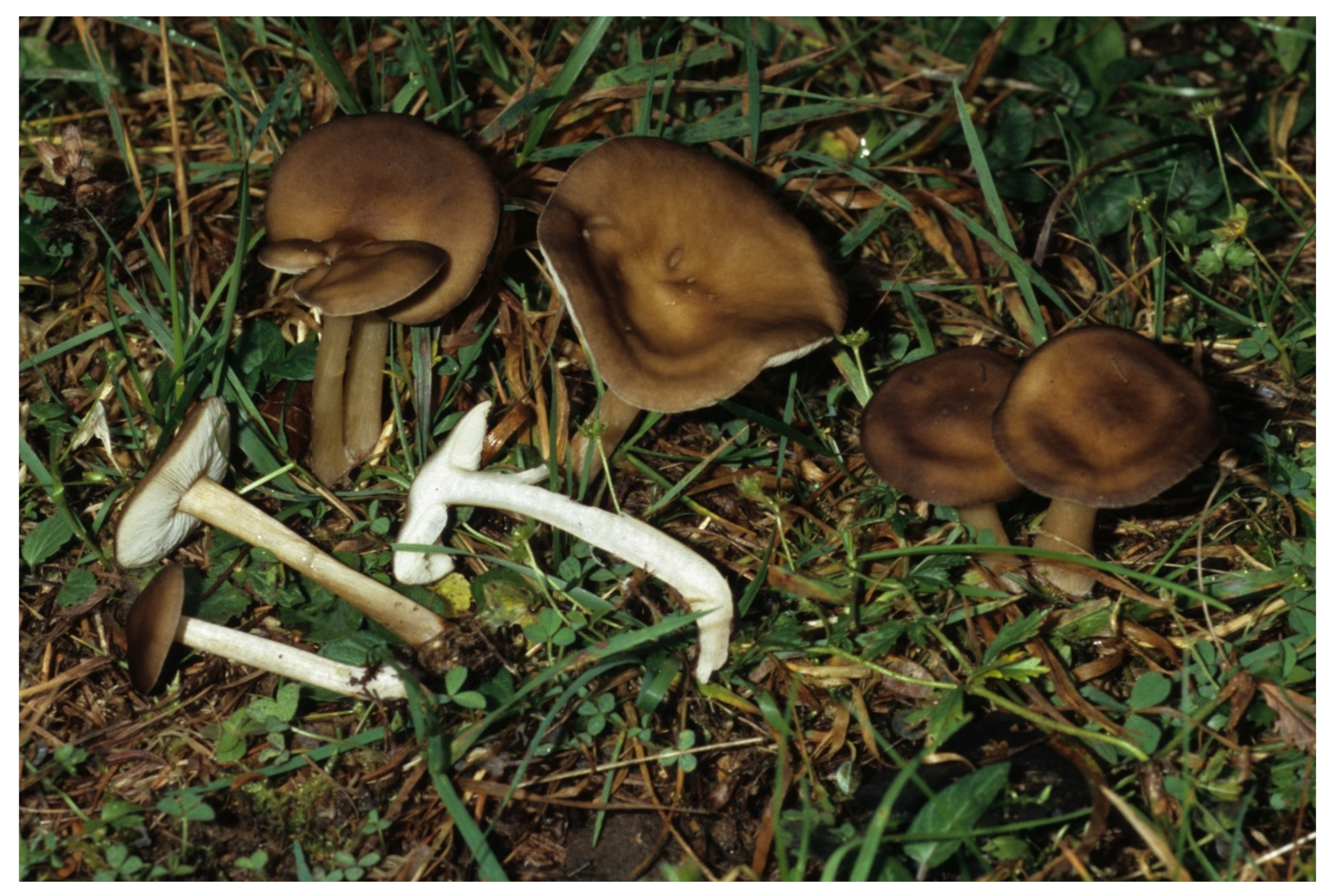
Melanoleuca fontenlae Para, Antonín, Ďuriška, Ševčíková & Tomšovský, sp. nov. (
Figure 5). MycoBank MB 838743 =
Melanoleuca pseudopaedida sensu Vizzini et al., 2011.
Diagnosis. Melanoleuca luteolosperma differs by slightly smaller basidiospores, (6.0)7.0–9.0 × (4.0)4.2–5.5(6.5) μm and by different sequences of ITS, tef1, and rpb2 genes.
Holotype. Italy, Emilia-Romagna Prov., Ravenna distr., Lido di Dante, 44°23′25″ N, 12°18′50″ E, 9 November 2000 leg. V. Antonín (BRNM 772194, GenBank/EMBL: MW491326—ITS, MW488158—tef1, MW488173—rpb2).
Etymology. Named “fontenlae” in honor of the Italian mycologist Roberto Fontenla, a longtime collaborator of R. Para in Melanoleuca studies.
Pileus 9–39 mm broad, broadly conical, or almost applanate, with indistinct or distinct, broad, conical, obtuse umbo at center, inflexed at margin, hygrophanous, margin smooth when young, later sometimes shortly striate, not pruinose, whitish gray, gray-brown, dark brown (5B2, 6C4, 7D3, 7E6–7, 7F5–6, 8E4–5), pallescent up to ochraceous brown to brown (6C–D4–6, 7D5–6), margin paler than center in young specimens. Lamellae moderately close, L = c. 30–65, l = 2–3, emarginate and attached with tooth, ± horizontal, not intervenose, whitish or cream-colored, then pale ochraceous (4A3) or grayish, with beige reflex, with concolorous, ± smooth, finely pubescent edge. Stipe longer than pileus width, 17–60 × 1.5–7 mm, cylindrical, slightly broadened at apex, clavate (up to 5 mm broad) at base, finely floccose-pubescent, especially at apex, entirely longitudinally fibrillose, lustrous, whitish to slightly brownish at apex, ochraceous yellowish to brownish (6C–D3–4, 7D–C4, 7D5) at center, dark gray-brown (7E–F4, 7E–F6, 8E–F3), with white or whitish basal mycelium. Context whitish, slightly grayish brownish under pileipellis, hollow, fibrillose, brown to dark brown in stipe base, with indistinct or slightly fungoid (earthy) smell and mild taste.
Basidiospores [240/10/10] (6.0)6.5–10 × (4.0)4.8–6.0(6.5) μm, average = 8.0 × 5.6 μm, E = (1.1)1.21–1.6(1.73), Q = 1.42, (broadly) ellipsoid, verruculose, warts mostly irregularly shaped and sized, sometimes up to 0.75 μm high, sometimes with rare ridges, amyloid. Basidia [37/6/6] 24–50 × 7.0–13 μm, four-, rarely two-spored, clavate. Basidioles 15–40 × 5.0–12 μm, clavate, subcylindrical. Cheilocystidia [82/10/9] (length × base width × apex width) 19–65 × 3.0–10 × 2.5–3.5 μm, urticoid of the brevipes- and exscissa-type, sometimes very rare or absent, basal part clavate, subfusoid, often irregular, apical part subulate, obtuse, rarely with two septa, thin-walled, with crystals or not. Marginal cells 15–40 × 4.0–10 μm, clavate, ± cylindrical, subfusoid, irregular or branched, thin-walled. Pleurocystidia absent. Trama hyphae ± cylindrical, fusoid to inflated, thin-walled, inamyloid, up to 15 μm wide. Pileipellis an ixocutis transient to ixo(sub)trichoderm at center; terminal cells adpressed to erect, cylindrical, (sub)fusoid, subclavate, obtuse, thin-walled, inamyloid, up to 50 × 10 μm wide, grayish brown in KOH. Stipitipellis a cutis of cylindrical, parallel, ± slightly thick-walled, inamyloid, up to 6.0 μm wide hyphae. Caulohymenium of (1) single or in groups, 21–50 × 6.0–10 μm, clavate, fusoid to cylindrical, sometimes narrowly cylindrical, 30–40 × 2.5–3.0 μm, with apical incrustation, with or without one septum, and (2) urticoid cystidia, 25–70 × 3.0–10 μm, with apical crystals or not, thin-walled; caulocystidia sometimes scattered to absent. Clamp connections absent.
Ecology. Growing on sandy soil and dunes; found in grass, on a path, under Pinus pinaster, Pinus maritima, Pinus pinea, Quercus ilex, Pyracantha, and Rubus and under Salix and Pinus and under Juniperus communis
Distribution. Known only to be from France, Italy, and Slovakia until now.
Additional specimens examined. France: Bais de Somme, 7 December 1981 leg. Mrs. Bergeron, det. M. Bon (PC, Romagnesi 81.253, as M. rasilis var. pseudoluscina).—Italy: Emilia-Romagna Prov., Ravenna distr., Pineta di San Vitale, Bardello, 10 November 2000 leg. V. Antonín (BRNM 825711).—Ibid., leg. M. Enderle (BRNM 825712).—Ibid., 9 November 2000 leg. V. Antonín and A. Hausknecht (BRNM 825713).—Ibid., Pineta Ramazzotti and Dunes di Lido di Dante, alt. –14 m a.s.l., 5 November 2007 leg. V. Antonín (BRNM 825714). — Venezia, Caorle, Valle Vecchia‒Brussa, 20 December 2014 leg. E. Campo (BRNM 825715).—Slovakia: Cerová vrchovina, Vlčia dolina, 27 October 2002 leg. K. Skokanová (SAV F-3825, SAV F-3822).—Ibid., 27 October 2004 leg. S. Adamčík (SAV F-3823, SAV F-3824).
Remarks. Melanoleuca fontenlae is characterized by small basidiomata with a gray-brown pileus pallescent up to ochraceous brown, whitish, or cream-colored, then pale ochraceous or grayish lamellae, a slightly clavate, brownish to dark gray-brown stipe, a brown to dark brown context in the stipe base, urticoid, but sometimes rare or even missing cheilocystidia and mostly well-developed caulohymenium.
Melanoleuca fontenlae is identical with
M. pseudopaedida sensu Vizzini et al. [
3]. However, the type specimen of
M. pseudopaedida, phylogenetically tallies to
M. luteolosperma. Therefore, we consider
M. pseudopaedida in the original sense as a synonymum of
M. luteolosperma.
Among phylogenetically close species, only Melanoleuca luteolosperma has similarly small basidiomata. It differs by slightly smaller basidiospores, (6.0)7.0–9.0 × (4.0)4.2–5.5(6.5) μm, average 7.7 × 5.1 μm. Melanoleuca paedida differs by a larger, 30–60 mm broad, ochraceous fawn to pale or dark gray-brown pileus; an only slightly darker context in the stipe base; and smaller basidiospores, 6.5–8.5 × 3.9–5.5 μm, average 7.3 × 5.1 μm; moreover, it constantly lacks a caulohymenium. Melanoleuca castaneofusca differs by larger basidiomata; a distinctly floccose to floccose-tomentose stipe at apex with an only brownish yellow tinged context in its base; and smaller basidiospores, 6.5–8.0 × 4.0–6.0 μm, average = 7.3 × 5.0 μm. Melanoleuca microcephala differs by a larger, (grayish) brown, beige-gray, or grayish pileus and consistently absent cheilocystidia and caulohymenium. Melanoleuca stridula (Fr.) Singer has a larger, 18–50 mm broad, rather dark (gray-)brown pileus; a distinctly bulbose stipe; slightly smaller basidiospores, 7.0–8.0(9.0) × 4.75–6.0 µm, average 7.9 × 5.4 µm; and consistently absent cheilocystidia and caulohymenium (our unpublished studies). The European collections of M. angelesiana have a larger, 25–80 mm broad pileus; an entirely floccose stipe with a pale context in its base; and constantly absent cheilocystidia and caulohymenium (our unpublished studies, see also notes above). Melanoleuca acystidiata has a brown pileus; a whitish, then light brown stipe; a white context in the stipe base; and a lack of cheilocystidia.
Melanoleuca robertiana Bon, Documents Mycologiques 20(79): 58, 1990.
Type revision (LIP 72092034, GenBank/EMBL: MW491341): Basidiospores 8.0–9.5(10) × 4.5–5.3 µm, average = 8.85 × 4.85 µm, E = 1.6–2.1, Q = 1.83, ellipsoid, oblong-ellipsoid, ellipsoid-fusoid, ornamentation of ± regular warts variable in size, ridges absent or very rare. Basidia 27–32 × 10–12 µm, four-spored, clavate or subfusoid. Basidioles 20–33 × 6.0–11 µm, clavate, subfusoid. Cheilocystidia 60–69 × 14–18 µm, fusoid or sublageniform, thin-walled with slightly thick-walled apex. Pleurocystidia absent. Trama hyphae cylindrical to ellipsoid, thin-walled, 5.0–15(20) µm wide, inamyloid. Pileipellis a cutis of cylindrical, thin-walled, 4.0–10 µm wide hyphae; terminal cells adpressed to suberect, cylindrical, obtuse, simple, rarely branched. Stipitipellis a cutis of cylindrical, parallel, 3.0–6.0 µm wide hyphae. Caulocystidia 28–41 × 7.0–12 µm, clavate, fusoid, subutriform, thin-walled; one macrocystidium seen, 44 × 13 µm. Clamp connections absent in all studied tissues.
Remarks. Bon [
28] described
Melanoleuca robertiana as an acystidiate taxon with basidiospores of (6.5)7–8.5(9) × (5)5.5–6(6.5) µm. The holotype was preserved in the Herbarium M. Bon, deposited in LIP (LIP 72092034). The majority of holotype material was not found in LIP, but a pocket with a small piece of holotype was glued to the original author’s sheet with a description of the holotype, consisting of roughly one-quarter of one small basidioma. Bon mentioned the absence of any cystidia and basidiospores of 6–7.5(8) × 5–6 µm on this description accompanying the holotype material. Macro- and microscopic characters of the
Melanoleuca robertiana published in the literature [
28,
29] indicates the possibility this species belonging to the
M. castaneofusca group. However, our holotype revision revealed the presence of 60–69 × 14–18 µm large cheilocystidia and basidiospores of 8.0–9.5(10) × 4.5–5.3 µm in size. The ITS sequencing of the holotype was successful, and ITS sequence agreed with that of macrocystidioid
M. pallidicutis Bresinsky holotype (TAAM 178616, MT270846) [
30] belonging to the subgenus
Melanoleuca. The obvious disagreements between these features of the studied piece of the type material and the characters described in the original protologue of
M. robertiana indicate that the original description and holotype specimen refer to two different taxa. It is almost certain that a mycologist as experienced as M. Bon described in the protologue a different collection than that which represents the type material of
M. robertiana. In the literature,
M. robertiana has always been considered an acystidiate species [
4,
10,
31,
32] (the last as synonym of
M. melaleuca). We are convinced that the material was mistakenly confused and a wrong basidioma was deposited in the herbarium envelope.
Melanoleuca pallidicutis is a taxon based on an unambiguous well-defined description and whose morphological characters in protologue match with that of the holotype and the DNA sequence supports its expected taxonomic position (Antonín et al. in prep.). On the basis of these facts, we considered the name
Melanoleuca robertiana Bon a
nomen confusum because there is a substantial conflict in crucial characters (the presence/absence and character of cystidia and spores size) between descriptions published in the protologue, other literature [
10,
28,
29], and the type specimen.
Vizzini et al. [
4] included, under the name
M. robertiana, the sequence of a fungus collected in Italy and with macro- and microscopic agreement with the original description by Bon [
28]. It is different from any other species of the
Urticocystis Boekhout [
2,
3,
4,
5]. We decided to describe it as a new species here.
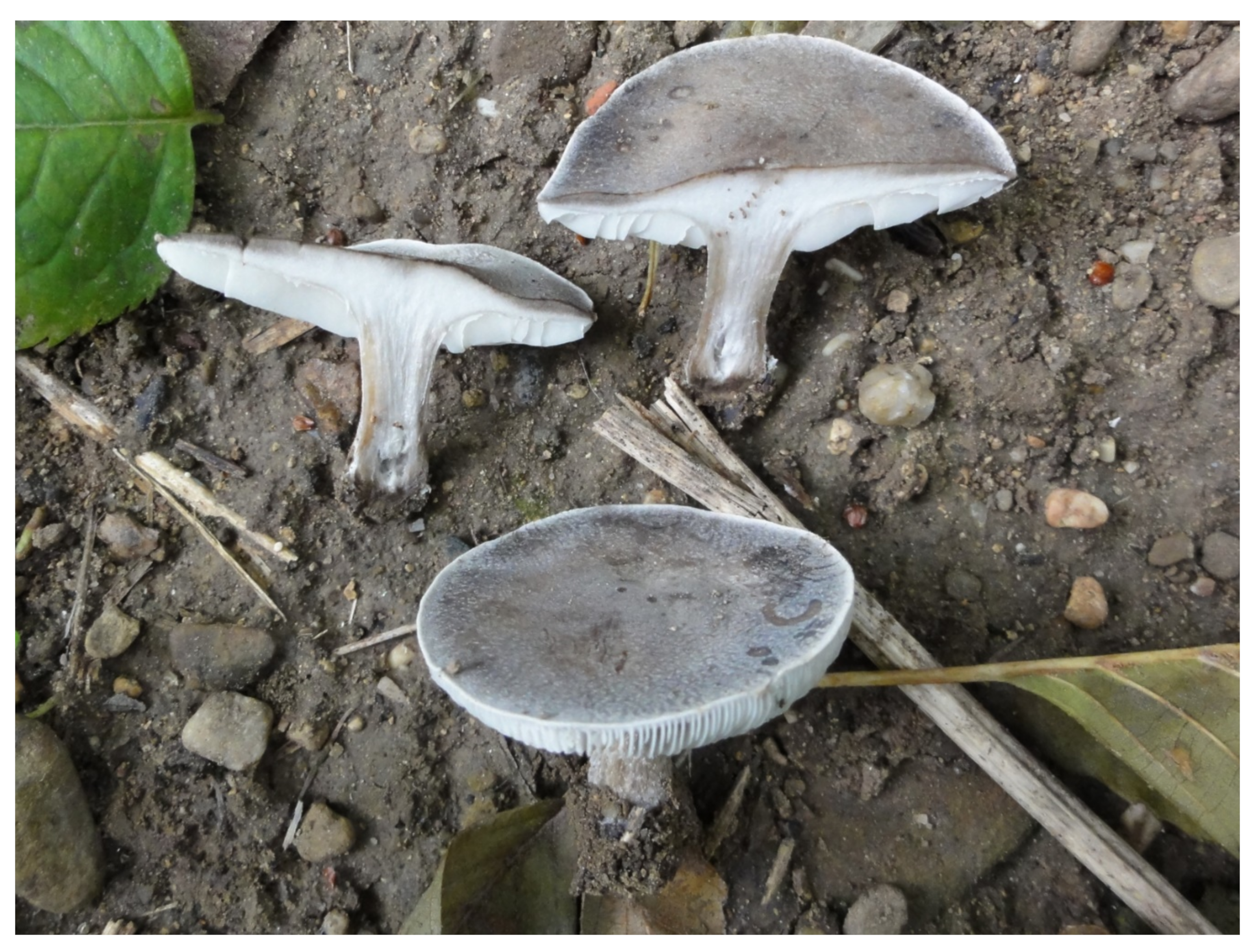
Melanoleuca acystidiata Para, Antonín, Ševčíková, Ďuriška & Tomšovský, sp. nov.
Figure 6 and
Figure 7.
Mycobank MB 838744
Diagnosis. It differs from M. microcephala by the pileus and stipe color and the white context in the stipe base, from M. paedida by the pileus color and slightly larger basidiospores (M. paedida 6.5–8.5 × 3.9–5.5 μm), and always absent cheilocystidia; from M. stridula by the pileus color and larger basidiospores (M. stridula 6.5–8(9) × 5–6 µm); and from M. fontenlae by the pileus and stipe color, white context in the stipe base, and absent cheilocystidia; all mentioned species also differ phylogenetically.
Holotype. Italy, South Tyrol, Bolzano Distr., St. Jacob in Val di Vizze, 46°54′03″ N, 11°28′14″ E, 1500 m a.s.l., numerous specimens on a grassy clearing of a Abies alba wood, 23 August 2007 leg. R. Para and R. Fontenla (ANC M0205, GenBank/EMBL: JN616462).
Etymology. Acystidiata—a species lacking cystidia.
Pileus 20–50 mm broad, from convex to irregularly flattened, with less distinct or absent umbo; margin inflexed; smooth, opaque, or silky-looking surface; hairless; slightly sticky when wet; finely velvety under lens; brown to dark brown (S60 Y70–99 M50, S80 Y10–99 M40) and sometimes almost black; sometimes paler or with ochre tinge (S50 Y40–50 M10, S40 Y 99 M20–40, S50 Y 99 M20–40); sometimes with discolored areas. Lamellae close, L = c. 60–70, l = 1–2 in between each lamella, emarginate, straight to slightly rounded, pure white to whitish or pale gray, sometimes darkening in age. Stipe usually (slightly) longer than pileus width, 30–70 × 2–7 mm, cylindrical, sometimes broadened at the base, surface glabrous, longitudinally fibrillose, sometimes twisted, in young basidiomata whitish, then light brown to brown with ochre tinge (S40 Y40–60 M10, S40 Y 99 M20–30), sometimes with indistinct whitish floccules. Context exiguous in the pileus; fibrous and tenacious in the stipe; white, whitish to light brown in the cortex; unchanging, with light herbaceous or fungoid smell and mild taste.
Basidiospores [96/3/3] (6.7)7.2–10(11) × 5–7.2(7.5) µm, average 8.2 × 6.1 µm, E = 1.19–1.64, E = 1.36; subglobose; broadly ellipsoid; ovoid; with ± large, regular, or irregular warts; ridges absent or rare; amyloid. Basidia [6/1/1] 29–37 × 11.5–13 µm, four-spored, clavate or subfusoid. Basidioles 12–35 × 5.0–13 µm, clavate, subcylindrical, subfusoid. Cheilocystidia and pleurocystidia not observed. Marginal cells 20–35 × 5.0–9.0 µm, clavate, (sub)cylindrical, mostly irregular, sometimes septate, thin-walled. Pileipellis acutis to ixocutis, consisting of cylindrical, thin-walled, up to 8.0 µm wide hyphae, inamyloid, smooth or with scattered simple to subcoralloid lateral projections; terminal cells adpressed to erect, cylindrical, narrowly fusoid or clavate, thin-walled, up to 10 µm wide; fusoid cystidioid elements rarely present. Stipitipellis a cutis of cylindrical, parallel, slightly thick-walled, up to 6.0 µm wide hyphae, inamyloid. Caulocystidia 22–40 × 7.0–12.5 µm, rare to frequent, in groups or isolated, clavate or cylindrical, sometimes rostrate. Clamp connections absent in all tissues.
Ecology. On soil in grass in montane spruce (Picea abies), fir (Abies alba), and larch (Larix decidua) stands on acidic soil of high elevation (≈1500–1900 m a.s.l.).
Distribution. Melanoleuca acystidiata has been found only in three montane localities in the north of Italy and Switzerland.
Additional specimens examined. Italy: Piemonte, Cuneo Distr., Bosco delle Navette di Ormea, alt. ≈1600 m a.s.l., 5 October 2015 leg. R Para and R. Fontenla (ANC M0233). — Switzerland: Davos, Schatzalp, alt. ≈1900 m, 25 June 2009 leg. J. Borovička (BRNM 772203).
Remarks. Melanoleuca acystidata is characterized by a more or less dark brown pileus; whitish lamellae; a stipe shorter or longer than the pileus width that is whitish when young, then light brown; a white context; subglobose; broadly ellipsoid or ovoid basidiospores, 7.2–9 × 6–7.2 µm in size; the absence of any hymenial cystidia; a pileipellis in the form of an (ixo)cutis with adpressed to erect terminal cells; and one type of clavate or cylindrical caulocystidia.
From the phylogenetically related species,
M. microcephala differs by a (grayish) brown, beige-gray, grayish or uniformly grayish brown pileus, a gray-brown to dark gray-brown stipe, and a dark (reddish) brown to black-brown context in the stipe base.
Melanoleuca paedida differs by an ochraceous fawn to pale or dark gray-brown pileus, slightly smaller basidiospores, 6.5–8.5 × 3.9–5.5 μm, average 7.3 × 5.1 μm, and occasional cheilocystidia.
Melanoleuca stridula has a uniformly rather dark, yellow-brown, (gray-)brown or dark brown pileus and smaller basidiospores, 6.5–8(9) × 5–6 µm, average 7.5 × 5.5 µm (our unpublished studies, [
31,
33,
34]).
Melanoleuca fontenlae has a whitish gray, gray-brown, dark brown pileus pallescent up to ochraceous brown or brown; an ochraceous yellowish to brownish, basally dark gray-brown stipe; a brown to dark brown context in the stipe base; and present cheilocystidia. The European collections of
M. angelesiana have a larger, 25–80 mm broad, uniformly silvery gray, gray-brown, brown, dark brown, and centrally up to black-brown pileus and a more robust (45–80 × 4–10 mm), entirely floccose gray-brown stipe (our unpublished studies). The syntype of
M. angelesiana, described from the Olympic Mts., Washington, USA, newly sequenced by us is more related to
M. acystidiata Bon than to
M. angelesiana identified by Vizzini et al. [
4], but both species are clearly different. Among other species without cystidia,
Melanoleuca brachyspora Harmaja differs by slightly smaller basidiospores, (6.5)7.0–9.0 × 4.5–6.5 μm, average = 7.5 × 5.5 μm. All above mentioned species also differ phylogenetically.
Melanoleuca castaneofusca Contu, Bulletin de la Fédération Mycologiques Dauphiné-Savoie 150: 41, 1998.
Figure 8.
Holotype. Italy: Sardinia, Orto Botanico di Cagliari, 3 November 1992 leg. M. Contu (CAG 921103-01!).
Pileus 24–100 mm broad, broadly conical to almost applanate, without umbo or with slightly distinct, broad, obtuse umbo at center, slightly reflexed towards margin, pileus cuticle exceeding the pileus margin, sometimes undulate, hygrophanous or not, smooth, glabrous or finely fibrillose, with silvery appearance in some pilei, not striate, uniformly dark gray-brown to brown (5YR3/1–2, 4/2; 7.5YR3/2–4, 4/1–2; 10YR4/1–4), pale brown (S30Y60M30) with darker brown center, without or with watery brownish stains. Lamellae moderately close, L = 40–60, l = 1–3(4), emarginate and adnexed with tooth, sinuate, rather narrow, cream-colored with a beige reflex (7.5YR8, 10YR8/1), edge concolorous, finely pubescent. Stipe usually shorter than pileus width, 12–70 × 4–10 mm, cylindrical, slightly broadened at apex, tapering towards the base, slightly clavate to clavate-bulbose (up to 15 mm) at base, distinctly floccose to floccose-tomentose at apex, otherwise longitudinally fibrillose, brownish to gray-brown or dark brown (4/2; 7.5YR3/2, 4/2; 10YR4/2–4), whitish or slightly brownish tinged at base; basal tomentum whitish. Context with slight fungoid smell and mild fungoid taste, white in pileus, brown beneath the pileipellis, fibrillose, whitish in stipe and with brownish yellow or pale orange tinge in the stipe base, in stipe cortex grayish.
Basidiospores [100/4/4] (6.0)6.5–8.0 × 4.0–6.0 μm, average = 7.1 × 4.9 μm, E = (1.20)1.28–1.70(1.75), Q = 1.47, (broadly) ellipsoid, ellipsoid-fusoid, with a suprahilar depression, with verruculose ornamentation, verruculae ± rounded, up to 0.5 μm high, variable in size, connections very rare to absent, amyloid. Basidia [28/4/4] 18–40 × 6.0–12 μm, four-spored, clavate to subfusoid. Basidioles 15–38 × 5.0–12 μm, clavate, subcylindrical, subfusoid. Cheilocystidia (length × base width × apex width) [31/3/3] 18–55 × 5.0–11 × 3.0–5.0 μm, urticoid, of the brevipes- and also exscissa-type, basal part fusoid, lageniform or clavate, septate or not, sometimes irregular, thin-walled, apical part subulate, obtuse with or without crystaliferous cap. Marginal cells 16–35 × 5.0–9.0 μm, fusoid, clavate, lageniform, irregular to (sub)coralloid, thin-walled. Pleurocystidia often absent, sometimes scattered, 30–40 × 5–10 μm, of the brevipes-type, with or without septum and apical incrustation. Trama hyphae cylindrical to inflated, thin-walled, inamyloid, up to 15 μm wide. Pileipellis an ixocutis, composed of cylindrical, ± thin-walled, gelatinized, up to 8.0 μm wide, smooth hyphae; terminal cells adpressed to (sub)erect, cylindrical, narrowly clavate, obtuse, thin-walled, with gray-vacuolar pigmentation. Stipitipellis a cutis of cylindrical, parallel, slightly thick-walled, up to 6.0 μm wide hyphae with gray-brown pigmentation. Caulocystidia (caulohymenium) of two types, (1) urticoid cystidia of the exscissa- or brevipes-type, 25–60 × 4.0–9.0 μm, both septate and not septate, thin-walled, with indistinct to distinct apical incrustation, and (2) clavate, subfusoid, or subcylindrical cells, 18–50 × 7.0–12 μm, thin-walled; cystidia of the type (1) may be very rare or even absent. Clamp connections absent.
Ecology. On soil under
Ulmus (BRNM 761900), in a greenhouse on a bare soil (BRNM 761901), on sandy soil under
Picea (LIP RC86021), in a flowerpot with
Mentha sp. in an urban apartment (SLO 1639), on composted soil in a cemetery [
35], and on soil and shredded bark mulch in a botanic garden (K(M) 92562); holotype on basic sandy soil near
Cactaceae in the botanic garden (CAG 921103-01). The species may be associated with commercial soil and compost substrates.
Distribution. This species is now confirmed from the Czech Republic, France, Great Britain (England), Italy, and Slovakia until now. However, we suppose it is more broadly distributed in similar habitats.
Specimens examined. Czech Republic: Osek near Hořovice, Vystrkov, 12 November 2012 leg. O. Jindřich (BRNM 761901).—France: Landrellec (Cotes du Nord), près Pleumeur-Boudon, 21 December 1986 leg. Reaudin, det. R. Courtecuisse (LIP RC86021, as M. brevipes).—Great Britain: England, Surrey, Kew, Royal Botanic Gardens, 9 October 2001 leg. A. Henrici (K(M) 92562, as M. turrita, a mixed collection with M. bataillei).—Italy: Sardinia, Orto Botanico di Cagliari, 3 November 1992 leg. M. Contu (CAG 921103-01, holotype).—Ravenna, Pineta di San Vitale, Casa vecchia, 8 November 2007 leg. M. Calderoni (BRNM 761900).—Slovakia: Podunajská nížina lowland, Bratislava, 28 October 2013 (SLO 1639).
Remarks. Melanoleuca castaneofusca, described from Sardinia [
36], is characterized by rather robust basidiomata with a dark-colored pileus; cream lamellae; an apically distinctly floccose to floccose-tomentose, brownish to gray-brown stipe; whitish and brownish yellow tinged context in the stipe base; rather small basidiospores; urticoid cheilocystidia of the brevipes- and exscissa-type; a pileipellis in the form of an ixocutis; and well-developed caulohymenium. This species produces its basidiomata in late autumn in habitats strongly influenced by humans or at completely artificial places (e.g., greenhouse, cemetery, botanic garden). In recent literature, only one collection (Great Britain, Surrey, Morden Cemetery, October 2012 leg. T. Brown) was published [
35]; it was identified on the basis of our sequences.
Among phylogenetically similar species, M. luteolosperma differs by smaller basidiomata (pileus 22–35 mm, stipe 50–55 × 3–4 mm), an ochraceous yellow or pale gray-brown stipe, and slightly larger basidiospores [7.0–9.0 × 5.0–5.5(6.0) μm]. Melanoleuca paedida differs by a smaller, 40–50 mm broad, ochraceous fawn to pale or dark gray-brown pileus and lacks a caulohymenium. Melanoleuca fontenlae differs by smaller basidiomata (pileus 9–39 mm broad, stipe 17–60 mm long), only finely floccose-pubescent stipe, a brown to dark brown context in the stipe base, and larger basidiospores [(6.0)6.5–10 × (4.0)4.8–6.0(6.5) μm, average = 8.0 × 5.6 μm].
Melanoleuca malenconii Bon is macroscopically somewhat similar but differs by pale to grayish yellow lamellae when mature; a slightly pruinose-pubescent to distinctly floccose, sometimes especially in lower part floccose-hairy stipe that is rarely subglabrous at apex; the presence of pleurocystidia; longer basidiospores (up to 10 μm); and sometimes also by relatively smaller, paler basidiomata [
2,
28].
Melanoleuca humilis (Pers.) Pat. is morphologically similar species different in its grayish or beige lamellae in mature basidiomata, a (dark) brown context in the stipe base, longer basidiospores (up to 10 μm), and often also a smaller pileus [
2].
≡ Agaricus luteolospermus Britzelm., Bericht der Naturhist Vereins in Augsburg 31: 160, 1894. ‒ Tricholoma luteolospermum (Britzelm.) Lapl., Dictionnaire Iconographique des Champignons Supérieurs: 540, 1894.
Lectotype. Britzelmayr, Bericht der Naturhistorischen Vereins in Augsburg 31: Table 647, 1894 [
37].
Epitype. Germany, vicinity of Augsburg, Wallenburg, Bergheim, Siebentischwald, Stadtberg n. Stätzling, Eurasburg, Peterhof, Wittelsbacher Park, 10 June 1962 leg. J. Stangl (M 0139486! [
37]).
= Melanoleuca pseudoluscina Bon, Documents Mycologiques 10(37–38): 89, 1979.
Holotype. France, Somme, Quend les Pins, fourrés de l’Hyppophaeion et Koelerion, October 1963 leg. M. Bon (LIP 31018!).
Epitype. Italy, Venezia, Pineta Ca’ Savio di Cavallino-Treporti, 12 December 1999 leg. E. Bizio (ANC M0192).
= Melanoleuca pseudopaedida Bon, Documents Mycologiques 20(79): 58, 1990.
Holotype. France, Quend les Pins (Somme), Panne de dunes, Salix et Pinus, 5 November 1983 leg. M. Bon (LIP, Bon 831105!).
Pileus 22–42(62) mm broad, convex with obtuse center, almost without umbo or applanate, depressed at center, inflexed to straight at margin, smooth or finely pruinose, glabrous, never translucently striate, uniformly pale ochraceous gray to gray (5A2–5B3, 5C1–5D1), grayish brown (6D–E4) or brown (6D7) with whitish very margin. Lamellae moderately close, L = c. 40–60, l = 2–3, emarginate and attached with a tooth, ventricose, broad, white to pale cream-colored (2–3A2), with concolorous, uneven, finely pubescent edge. Stipe shorter or longer than pileus width, 20–55 × 3–7 mm, cylindrical, slightly broadened at apex, slightly broadened or attenuated and shortly radicating at base, finely longitudinally fibrillose, finely floccose-pubescent at apex, whitish at apex, pale ochraceous yellow (3A2) or pale gray-brown (±6C3) otherwise. Context whitish in pileus, whitish in stipe apex and (dark) brown (±6E4) in base, with indistinct smell and mild to bitterish taste.
Basidiospores [180/8/8] (6.0)7.0–9.0 × (4.0)4.2–5.5(6.5) μm, average 7.7 × 5.1 μm, E = (1.20)1.26–1.74(1.8), Q = 1.50, broadly ellipsoid, broadly ellipsoid, ellipsoid-fusoid, subovoid, verruculose, warts mostly irregular in shape and variable in size, up to 0.75 μm high, amyloid, ridges rare. Basidia [38/6/6] 26–46 × 8.0–12 μm, four-spored, clavate, subfusoid. Basidioles (12) 17–40 × 4.0–12 μm, clavate, subfusoid, cylindrical. Cheilocystidia [80/8/8] (length × base width × apex width) 22–66 × 3.0–10 × 3.0–4.5 μm, urticoid, of the exscissa- and brevipes-type, basal part clavate, subvesiculose, subfusoid, sometimes irregular, apex subulate or (sub)cylindrical, thin- to slightly thick-walled, muricate or not. Marginal cells 12–24 × (4.0)8.0–12 μm, clavate, vesiculose, sometimes two- or three-celled, thin-walled. Pleurocystidia absent. Trama hyphae cylindrical or subinflated, thin-walled, inamyloid, 3.0–15 μm wide. Pileipellis an (ixo)cutis, sometimes transient to subtrichoderm, composed of radially arranged, cylindrical, thin-walled, inamyloid, 3.0–8.0 μm wide hyphae; terminal cells often cystidioloid, adpressed to erect, cylindrical to clavate or fusoid, sometimes irregular to branched, thin-walled, obtuse, up to 8.0 μm wide, grayish brown in KOH. Stipitipellis a cutis of cylindrical, parallel, slightly thick-walled, inamyloid, 3.0–6.0 μm wide hyphae. Caulohymenium of (1) 16–70 × 2.0–11 μm, clavate, cylindrical, thin-walled, sometimes uniseptate cells; (2) urticoid cystidia, 24–64 × 4.0–10 μm, similar to cheilocystidia; and (3) caulobasidia, four-spored. Clamp connections absent.
Ecology. On soil, under Picea abies and Fagus sylvatica in a montane forest and on the riverbank under Populus, Sambucus, and Alnus (Slovakia), on the riverbank among grass, Dryas, and Picea (Slovenia, along the path in decaying remnants of grasses; under Juglans regia, Rosa, and Clematis (Italy); sandy soil under Populus (Italy); under Cistus (Italy); in a thermophilic forest stand (Czech Republic); and in a semi-dry grassland with Pinus (Germany).
Distribution. This species is widely distributed in Europe. Its occurrence is confirmed from the Czech Republic, Germany, Italy, Slovakia, and Slovenia.
Specimens examined. Czech Republic: Bohemian Karst, Koda, 28 October 2009 leg. J. Burel and O. Jindřich (BRNM 817820).—France: Somme, Quend les Pins, thickets of Hyppophaeion and Koelerion, October 1963 leg. M. Bon (LIP 31018, holotype of M. pseudoluscina).—Quend les Pins (Somme), dunes, Salix and Pinus, 5 November 1983 leg. M. Bon (LIP, Bon 831105, holotype of M. pseudopaedida).—Germany: vicinity of Augsburg, Wallenburg, Bergheim, Siebentischwald, Stadtberg n. Stätzling, Eurasburg, Peterhof, Wittelsbacher Park, 10 June 1962 leg. J. Stangl (M 0139486, epitype).—Lahr-Sulz, Pinus, 20 June 1998 leg. G. Saar (BRNM 772190).—Italy: Calabria, Pollino National Park, Lungro, Piano di Campolungo, 8 October 2019 leg. H. Ševčíková (BRNM 826043).—Monti Sibillini,
Altino, 22 October 2010 leg. V. Antonín (BRNM 817785).—Bibione, Faro, 8 December 2014 leg. E. Campo (817784).—Venezia, Pineta Ca’ Savio di Cavallino-Treporti, 12 December 1999 leg. E. Bizio (ANC M0192, epitype of M. pseudoluscina).—Ferrara, loc. Bosco Spada di Codigoro (FE), 14 November 2008 leg. G. Consiglio and A. Gennari (ANC M0194, as M. pseudoluscina).—Venezia, Giardini Ca’ Bianca, 14 November 1993 leg. E. Bizio (ANC M0191, as M. pseudoluscina).—Venezia, loc. Pineta Ca’ Savio di Cavallino-Treporti, 20 December 1998 leg. L. Levorato and C. Losi (ANC M193, as M. pseudoluscina).—Belluno, Caviola, Bosco Di Biasio di Falcade, 16 August 1996 leg. E. Bizio (ANC M0195, as M. pseudoluscina). — Slovakia: Velká Fatra Mts., Lubochňa, Lubochňanská dolina, Kundračka National Nature Reserve, alt. 820–1280 m a.s.l., 31 August 2002 leg. V. Kabát and V. Antonín (BRNM 761907).—Považský Inovec, Moravany nad Váhom, Výtoky, 29 September 2012 leg. I. Stach (SLO 1591).—Biele Karpaty, Skalka nad Váhom, near river Súčanka, 10 October 2013 leg. O. Ďuriška (SLO 1632).—Devínska Kobyla, Fialková dolina valley, 28 November 2019 leg. O. Ďuriška (SLO 2515).—Slovenia: Julians Alps, Triglav National Park, Zadnja Trenta, Upper Soča valley, alt. 980 m a.s.l., 10 October 2001 leg. G. Podgornik (BRNM 772201).
Remarks. Melanoleuca luteolosperma is an extremely variable species in terms of colors, characterized by rather small basidiomata with a uniformly pale ochraceous gray to gray or grayish brown pileus; pale cream lamellae; a cylindrical, basally slightly broadened or attenuated stipe that is finely floccose-pubescent only at apex and pale ochraceous yellow or pale gray brown; a pale brown context; moderately large basidiospores ((6.0)7.0–9.0 × (4.0)4.2–5.5(6.5) μm), varying from broadly ellipsoid (Q = 1.38) to ellipsoid (Q = 1.59); urticoid cheilocystidia of both the exscissa- and brevipes-type; a pileipellis often forming cystidioloid terminal cells; and mostly well-developed caulohymenium. The collection from Italy (Altino, BRNM 817785) differs by smaller basidiospores [6.5–7.5 × 4.5–5.5(6.0) μm].
Métrod [
33] and Bon [
10] considered
M. luteolosperma a species with macrocystidia. However, Fontenla and Para [
37] selected the epitype from the material collected in the original locality [
38]. This epitype has urticoid cheilocystidia.
M. luteolosperma was described as having a yellowish white spore print and whitish to yellowish basidiospores; its lamellae should be white when young, or whitish or brownish white [
39]. The sequence of this epitype is identical to several recent collections with white, whitish, or only slightly cream lamellae. Therefore, we conclude that
M. luteolosperma is a fungus with a broader variability of the lamellae color and with urticoid cystidia.
The sequence identified as
M. pseudoluscina (JN616455) from Italy [
4] was grouped among those of
M. luteolosperma. Unfortunately, the sequencing of the holotype specimen of
M. pseudoluscina failed. The holotype specimen (LIP 31018) is described as having a gray, often initially pale, then gray-brown or fuligineous and finaly black-gray (“atro-ardosiacus”) pileus, and a brown (“aerino-fuscus”) or fuligineo-gray (“fuligonoso-ardosiacus”) stipe [
40]; microscopically, except for slightly different basidiospores [7.0–9.0(9.5) × 5.2–6.0(6.5) μm, average 8.1 × 5.7 μm], it agrees with the description of
M. luteolosperma above. The holotype specimen of
M. pseudopaedida is phylogenetically identical with
M. luteolosperma, but different from
M. pseudopaedida sensu Vizzini et al. [
3], which is described as a new species
M. fontenlae above.
Among phylogenetically close species, Melanoleuca castaneofusca differs by larger basidiomata, a darker dark gray-brown to brown pileus, a darker brownish to gray-brown or dark brown stipe, and slightly smaller basidiospores (6.5–8.0 × 4.0–6.0 μm). Melanoleuca paedida differs by a slightly larger, 30–60 mm broad, ochraceous fawn to pale or dark gray-brown pileus; a stipe that is brownish, ochraceous orange, or concolorous with the pileus; slightly different basidiospores; and a lack of a caulohymenium. Melanoleuca fontenlae differs by a darker pileus and stipe and larger basidiospores [(6.0)6.5–10 × (4.0)4.8–6.0(6.5) μm, average = 8.0 × 5.6 μm].
The European collections of
M. angelesiana A.H. Sm. are macroscopically similar to
M. luteolosperma. It differs by larger basidiomata (pileus 25–80 mm, stipe 45–80 × 4–10 mm); a whitish context in the stipe base; larger basidiospores, 7.0–10 × (4.5)5.0–6.5(7.0) µm; and large basidia and basidioles (30–55 × 10–12 μm) (our unpublished studies). However, this species was originally described in North America, and the identity of European collections in terms of the work of Vizzini et al. [
3] and American specimens is questionable.
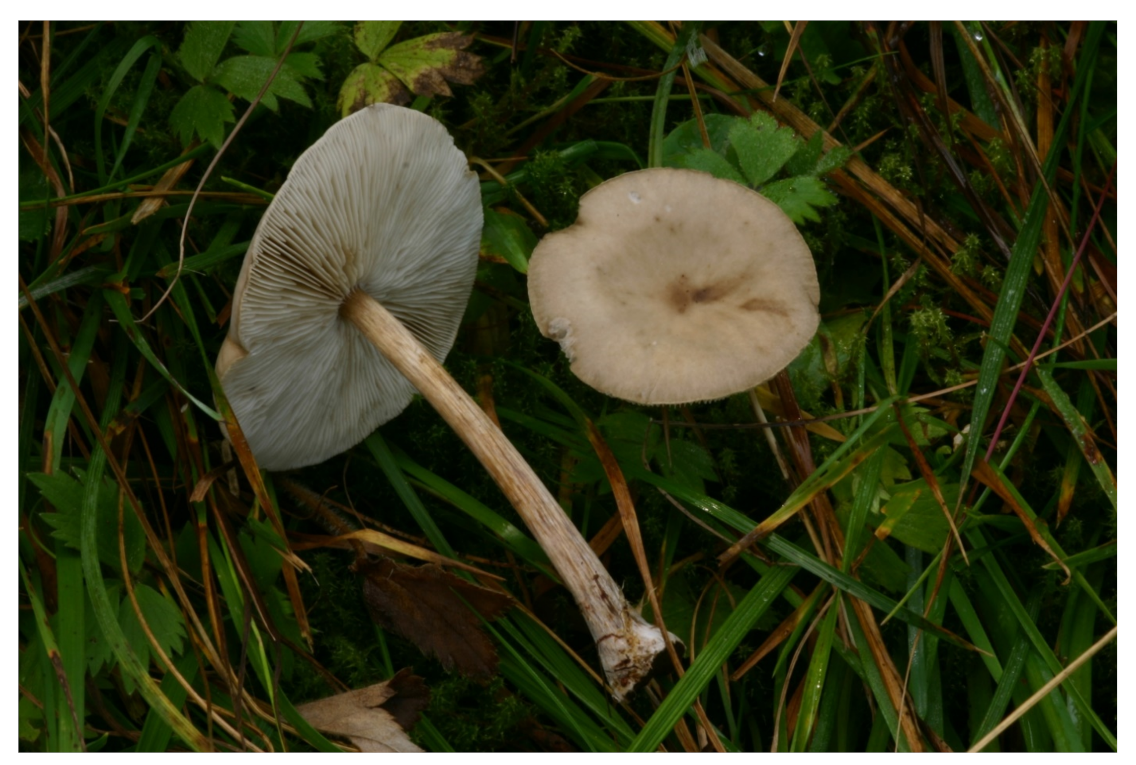
Melanoleuca microcephala (P. Karst.) Singer, Cavanillesia 7: 123, 1935.
Figure 12.
≡ Tricholoma microcephalum P. Karst., Hedwigia 20 (12): 177, 1881.
Lectotype. Finland, Tavastia australis, Tammela, Mustiala, 26 August 1881 leg. et det. P.A. Karsten (H; Herbarium Petter Adolf Karsten, no. 1604, as Tricholoma microcephalus, designated here, MycoBank MBT 395959).
Pileus 18–60 mm broad; broadly low convex-conical; conical to applanate and centrally depressed with low, broad, and less distinct umbo; inflexed to involute at margin, with pileipellis slightly projecting beyond the margin; hygrophanous; sometimes slightly translucently striate at very margin; smooth, glabrous, or slightly aeriferous at margin; finger test + or 0; (grayish) brown (7–8F3–6, 6E–F4) at center; beige-gray or grayish (6B–E2–3, 6D3–4), otherwise uniformly grayish brown (6C–D–E4–5). Lamellae moderately close, L = c. 30–60, l = 2–4, emarginate and attached to shortly decurrent with tooth, sinuate when young, up to ≈5 mm broad, whitish to pale cream-colored with beige reflex, with concolorous, finely pubescent edge. Stipe usually longer than pileus width, 22–105 × 2–5.5 mm; cylindrical; clavate to subbulbose towards base (9–12 mm); slightly broadened at apex; finely pruinose or floccose, especially at apex; longitudinally fibrillose or almost fibrillose-squamulose; whitish to pale brownish at apex; pale yellowish with ochraceous tinge (near 4A3); gray-brown (6F2–5) to dark gray-brown towards base; fibrils often darker than ground color when moist—therefore, the stipe gives a mottled appearance; basal tomentum white. Context whitish in pileus, brownish under the pileipellis, fibrillose and pale to dark (reddish)brown (6–7F6) to black-brown in stipe base, brownish in stipe cortex, without smell or with a slight earthy smell and mild taste.
Basidiospores 7.0–9.5(10) × 5.0–7.0 μm, average = 8.3 × 5.8 μm, E = 1.21–1.67, Q = 1.42, broadly ellipsoid, ellipsoid, ellipsoid-fusoid, obovoid, thin- to slightly thick-walled, ornamentation verrucose, sometimes with rare ridges, warts variable in shape and size, round or irregular, close to moderately close, amyloid. Basidia 28–37 × 9.0–13 μm; four-, very rarely two-spored; clavate. Basidioles 12–35 × 5.0–13 μm, clavate, subcylindrical, subfusoid. Cheilo- and pleurocystidia observed. Marginal cells 17–28 × (3.5)5.0–11 μm, cylindrical, clavate, subfusoid, utriform, often irregular, thin-walled. Trama hyphae cylindrical to subinflated, thin-walled, inamyloid, 3.0–15 μm wide. Pileipellis an ixocutis (margin) to ixosubtrichoderm (centre), composed of radially arranged, ± cylindrical, thin-walled, up to 10 μm wide hyphae with often grayish to gray-brown pigmentation; terminal cells adpressed to (sub)erect, up to ≈50 × 3.0–10 μm, clavate, cylindrical, subfusoid, obtuse, thin-walled; pale grayish yellowish in KOH. Stipitipellis a cutis of cylindrical, parallel, ± slightly thick-walled, 3.0–6.0 μm wide hyphae. Caulocystidia in groups, 18–62 × 5.0–15 μm, cylindrical, clavate, subfusoid, thin-walled. Clamp connections absent.
Ecology. On soil in the montane and alpine belt, on a pasture with scattered Picea, in an alpine meadow, in mosses and vegetation along the montane stream, in grass in spruce forest, and along the forest path. It seems to be a montane calciphilous species.
Distribution. It is known from the montane and alpine belt on altitudes of ≈1500 m or more a.s.l. It is only confirmed from Finland (lectotype), Italy, and Slovakia until now.
Additional specimens examined. Italy: South Tyrol, Schlern-Rosengarten Naturpark (Sciliar-Catinaccio Nature Park), Tiers (Tires), alt. 1647 m a.s.l., 4 September 2013 leg. V. Antonín 13.212 (BRNM 817786). — South Tyrol, Prato Nuovo (Neuwies), Franzenshöhe, N 46°31′55″, E 010°28′46″, alt. 2150 m a.s.l., 30 July 2018 leg. B. Dima (BRNM 809300). — South Tyrol, Sulda (Solden), N 46°31′24″, E 010°35′17″, alt. 1864 m a.s.l., 31 July 2018 leg. J. Hrabáková (BRNM 817789). — South Tyrol, Franadega di Dobbiaco (BZ), alt. 1600 m a.s.l., 21 July 2005 leg. W. Tommasi (ANC M0196/JN616449 and herb. R. Para 050721-02, originally as M. stepposa). — Slovakia: Velká Fatra Mts., Ružomberok, Podsuchá part, Smrekovica, Skalná Alpa National Nature Reserve, alt. 1290–1320 m a.s.l., 23 September 2009 leg. V. Antonín (BRNM 817787 and 761890). — Velká Fatra Mts., Ružomberok, Podsuchá part, Smrekovica, protecting belt of the Skalná Alpa National Nature Reserve, alt. 1300–1367 m a.s.l., 24 September 2009 leg. V. Antonín (BRNM 817788). — Belianske Tatry Mts., Tatranská Kotlina, Skalné vráta, alt. ≈1450–1950 m a.s.l., 5 September 2001 leg. I. Milan (BRNM 772196).
Remarks.
Melanoleuca microcephala is characterized by moderately large basidiomata with a (grayish) brown, beige-gray, grayish, or uniformly grayish brown pileus; whitish to pale cream lamellae; a gray-brown to dark gray-brown stipe; a dark (reddish)brown to black-brown context in the stipe base and the absence of cheilo- and pleurocystidia and the absence of caulohymenium. Two cystidioid elements, 35–36 × 7.0–8.0 μm, on the lamellar edge were found only in the Italian collection (Italy, Tiers, BRNM 817786). However, they were not typically urticoid cells, but structures such as a transient form between cystidia and marginal cells. An atypical pale orangish yellow coloring of the stipe after touching appeared in the Slovak collection (Ružomberok, BRNM 817788). Caroti et al. [
41] published a collection of
M. microcephala with a small, 15–20 mm broad, dark brown pileus with a white outermost margin and scattered urticoid cheilo- and pleurocystidia. However, this collection was not confirmed by phylogenetic studies and its identity is unclear.
In comparison with phylogenetically similar species,
Melanoleuca paedida differs by an ochraceous fawn to pale or dark gray-brown pileus, a stipe of the same length or shorter than the pileus diameter, a slightly darker context in the stipe base, and slightly smaller basidiospores; the collection with cystidia as well as the acystidiate collection are known in this species.
Melanoleuca stridula has a uniformly rather dark, yellow-brown, (gray-)brown, or dark brown pileus and smaller basidiospores, 6.5–8(9) × 5–6 µm, average 7.5 × 5.5 µm (our unpublished studies, [
31,
33,
34]).
Melanoleuca acystidiata has a dark brown pileus; a whitish, then light brown stipe; and a dark brown context in the stipe base. Among other species without cystidia,
M. brachyspora Harmaja differs by a differently colored, brownish gray or brown to dark brown pileus; a white context in the stipe base; and slightly smaller basidiospores, (6.5)7.0–9.0 × 4.5–6.5 μm, average = 7.5 × 5.5 μm. The European collections of
M. angelesiana differs by a whitish context in the stipe base and large basidia and basidioles (30–55 × 10–12 μm) (our unpublished studies, see also above).
Melanoleuca paedida (Fr.) Kühner & Maire, Bulletin trimestriel de la Société mycologique de France 50: 18, 1934.
Figure 13.
≡ Agaricus paedidus Fr., Epicrisis Systematis Mycologici: 53, 1838. ‒ Gyrophila paedida (Fr.) Quél., Enchiridion Fungorum: 18, 1886. ‒ Tricholoma paedidum (Fr.) Quél., Mémoires de la Société d’Émulation de Montbéliard, Série 2, 5: 341, 1873.
Neotype. Fries, Icones selectae Hymenomycetum nondum delineatorum, Table 46, Figure 1, 1867–1884 (designated here, MycoBank MBT 395960).
Pileus 30–60 mm broad, convex, soon flattened, then with depressed center, with absent or very reduced umbo and entire margin, glabrous (silky under lens, positive finger test), dry, opaque, ochraceous fawn to pale or dark gray-brown, with darker center. Lamellae moderately close to close, with numerous lamellulae of various lengths, emarginate and attached with tooth, high, straight, whitish or brownish gray, with concolorous edge. Stipe shorter than 30 × 6 mm or 60 × 4–8 mm, shorter or of the same length as the pileus width, cylindrical, enlarged or clavate at the base, with barely pruinose surface at the apex only, smooth or striate, brownish, ochraceous orange or concolorous with the pileus, with widespread white mycelial felt at the base. Context white or whitish, immutable, slightly darker in the stipe base, brownish also in the cortex of the stem and between the pileus and the lamellae, with pleasant herbaceous smell.
Basidiospores 6.5–8.5 × 3.9–5.5 μm, average 7.3 × 5.1 μm, E = 1.20–1.76, Q = 1.43, ellipsoid or subglobose, with small to medium sized, not close, amyloid warts. Basidia 26–40 × 9–10 μm, clavate, with long and narrow base, four- and two-spored. Cheilocystidia 50–74 × 5–10 μm, very numerous, urticoid. Pleurocystidia numerous, similar to cheilocystidia. Cheilo- and pleurocystidia may sometimes absent. Marginal cells 20–55 × 5–10 μm, scattered, clavate, twisted. Trama hyphae parallel, cylindrical. Pileipellis an ixocutis of interwoven hyphae, 3–4 µm wide, with a very thin gelatinous layer, with lemon-yellow intracellular pigment in superficial hyphae, yellowish-brown in underlying hyphae. Stipitipellis of parallel hyphae, with rare emerging hairs of variable shape. Caulocystidia absent. Clamp connections absent.
Ecology. On soil in Cedrus atlantica, Cedrus brevifolia, and Cedrus libani litter (Calabria) and in the grass under Pinus sp. (Lombardia).
Distribution. Recently confirmed only from Italy, but its distribution may be broader in Europe.
Specimens examined. Italy: Calabria, Colamauci di Celico (CS), alt. 1200 m a.s.l., 16 November 2009 leg. C. Lavorato (ANC M0189, JN616452). — Lombardia, lago Cancano di Valfurva (SO), alt. 1900 m a.s.l., 7 September 2001 leg. E. Carassai (herb. R. Para 010907-02).
Remarks.
Melanoleuca paedida is characterized by an ochraceous fawn to pale or dark gray-brown pileus with darker center; whitish or brownish gray lamellae; a stipe shorter or of the same length as the pileus diameter; brownish, ochraceous orange, or concolorous with the pileus; a slightly darker context in the stipe base, moderately large, ellipsoid, or subglobose basidiospores; and lacking caulocystidia. Cheilo- and pleurocystidia are either present (ANC M0189) or absent (010907-02). The variability of the latter character requires further study. Therefore, the epitype is not proposed here. The identity of this species is based on the studies by Vizzini et al. [
4].
Among phylogenetically similar species, Melanoleuca castaneofusca differs by a larger, uniformly dark gray-brown to brown pileus with watery brownish stains, brownish to gray-brown or dark brown stipe that is a distinctly floccose to floccose-tomentose at apex, constantly present cheilocystidia, and well-developed caulohymenium. Melanoleuca luteolosperma has a smaller, 22–35 mm broad pileus with a whitish very margin, a pale ochraceous yellow or pale gray-brown stipe, slightly smaller basidiospores, constantly present cheilocystidia, and a well-developed caulohymenium. Melanoleuca fontenlae differs by a brown to dark brown context in the stipe base and larger basidiospores [(6.0)6.5–10 × (4.0)4.8–6.0(6.5) μm, average = 8.0 × 5.6 μm], and it usually has a well-developed caulohymenium or at least caulocystidia. Melanoleuca stridula has a longer stipe than the pileus dimeter, slightly lager basidiospores [7.0–8.5(9.0) × 4.7–6.0 μm, average = 7.9 × 5.4 μm], constantly absent cheilocystidia, and a developed caulohymenium (our unpublished studies). Melanoleuca acystidiata differs by a dark brown pileus; a whitish, then light brown stipe; a white context in the stipe base; slightly larger basidiospores, 7.2–9 × 6–7.2 µm, average 8.6 × 6.3 µm; and always absent cheilocystidia.
European collections of M. angelesiana are also morphologically similar. They differ by a larger, 25–80 mm broad, uniformly silvery gray, gray-brown, brown, dark brown, and centrally up to black-brown pileus; a more robust (45–80 × 4–10 mm), gray-brown stipe; larger basidiospores [7.0–10 × (4.5)5.0–6.5(7.0) μm, average = 8.5 × 5.8 μm]; and consistently absent cheilocystidia and clavate caulocystidia.


 ,
, 


